

In Vivo 3-Dimensional Dynamic Evaluation of Shoulder Kinematics After the Latarjet Procedure: Comparison With the Contralateral Healthy Shoulder
- PMID: 38486807
- PMCID: PMC10938626
- DOI: 10.1177/23259671241226909
In Vivo 3-Dimensional Dynamic Evaluation of Shoulder Kinematics After the Latarjet Procedure: Comparison With the Contralateral Healthy Shoulder
Abstract
Background: Researchers have attempted to understand the underlying mechanism of the Latarjet procedure; however, its effects on shoulder kinematics have not been well studied.
Purpose/hypothesis: The purpose was to analyze shoulder kinematics after the Latarjet procedure. It was hypothesized that the nonanatomic transfer of the coracoid process during the procedure would affect normal shoulder kinematics.
Study design: Controlled laboratory study.
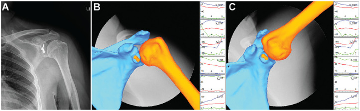
Methods: The study included 10 patients (age range, 20-52 years) who underwent the modified Latarjet procedure between June 2016 and November 2021. Computed tomography and fluoroscopy were conducted on both shoulder joints of all patients, and 3-dimensional models were reconstructed. The 3-dimensional coordinates were encoded on the reconstructed models, and shoulder kinematics were analyzed through a 3-dimensional-2-dimensional model-image registration technique. Scapular rotation parameters (scapular upward rotation, posterior tilt, external rotation, and scapulohumeral rhythm) were compared between the Latarjet and the nonsurgical contralateral sides during humeral abduction, as was anteroposterior (AP) translation relative to the glenoid center during active humeral external rotation.
Results: The Latarjet side displayed significantly higher values of scapular upward rotation at higher degrees of humeral elevation (130°, 140°, and 150°) compared with the nonsurgical side (P = .027). Posterior tilt, external rotation, and scapulohumeral rhythm were not significantly different between sides. AP translation at maximal humeral rotation was not significantly different between sides (Latarjet, -0.06 ± 5.73 mm vs nonsurgical, 5.33 ± 1.60 mm; P = .28). Interestingly, on the Latarjet side, AP translation increased until 40° of humeral rotation (4.27 ± 4.64 mm) but began to decrease from 50° of humeral rotation.
Conclusion: The Latarjet side demonstrated significant changes in scapular upward rotation during higher degrees of humeral elevation compared with the contralateral shoulder. Posterior movement of the humeral head at >50° of humeral rotation could be the desired effect of anterior stabilization; however, researchers should evaluate long-term complications such as osteoarthritis.
Clinical relevance: Analysis of shoulder kinematics after the Latarjet procedure could provide information regarding long-term outcomes and whether the procedure would affect the daily activities of patients.
Keywords: 3D-2D model-image registration technique; Latarjet; scapulohumeral rhythm; shoulder kinematics.
© The Author(s) 2024.
Conflict of interest statement
One or more of the authors has declared the following potential conflict of interest or source of funding: Grant support was received from the National Research Foundation of Korea funded by the Korean government (NRF-2021R1C1C1003481 to H.H.). AOSSM checks author disclosures against the Open Payments Database (OPD). AOSSM has not conducted an independent investigation on the OPD and disclaims any liability or responsibility relating thereto. Ethical approval for this study was obtained from Yonsei University Wonju Severance Christian Hospital (ref No. CR322005).
Figures


Similar articles
-
Kinematic stabilization after the Latarjet procedure: beyond the triple blocking effect.J Shoulder Elbow Surg. 2024 Oct;33(10):e547-e558. doi: 10.1016/j.jse.2024.02.022. Epub 2024 Mar 26. J Shoulder Elbow Surg. 2024. PMID: 38548097
-
Evaluation of three-dimensional in vivo scapular kinematics and scapulohumeral rhythm between shoulders with a clavicle hook plate and contralateral healthy shoulders.Int Orthop. 2019 Feb;43(2):379-386. doi: 10.1007/s00264-018-4003-y. Epub 2018 Jun 8. Int Orthop. 2019. PMID: 29948011
-
The Effects of Latarjet Reconstruction on Glenohumeral Kinematics in the Presence of Combined Bony Defects: A Cadaveric Model.Am J Sports Med. 2016 Jul;44(7):1818-24. doi: 10.1177/0363546516635651. Epub 2016 Apr 15. Am J Sports Med. 2016. PMID: 27159305
-
Shoulder muscle activity and function in common shoulder rehabilitation exercises.Sports Med. 2009;39(8):663-85. doi: 10.2165/00007256-200939080-00004. Sports Med. 2009. PMID: 19769415 Review.
-
Arthroscopic Bankart repair with remplissage versus Latarjet procedure for management of engaging Hill-Sachs lesions with subcritical glenoid bone loss in traumatic anterior shoulder instability: a systematic review and meta-analysis.J Shoulder Elbow Surg. 2020 Oct;29(10):2163-2174. doi: 10.1016/j.jse.2020.04.032. Epub 2020 Jun 9. J Shoulder Elbow Surg. 2020. PMID: 32807370
Cited by
-
Dynamic anterior stabilization for recurrent anterior shoulder instability improves postoperative patient-reported outcomes without restricting shoulder range of motion: a meta-analysis.Int Orthop. 2025 Aug;49(8):1931-1941. doi: 10.1007/s00264-025-06581-6. Epub 2025 Jun 16. Int Orthop. 2025. PMID: 40522491
-
Scapular kinematics variability in individuals with and without rotator cuff-related shoulder pain: A systematic review with multilevel meta-regression.Braz J Phys Ther. 2025 Aug 21;29(6):101261. doi: 10.1016/j.bjpt.2025.101261. Online ahead of print. Braz J Phys Ther. 2025. PMID: 40845626 Free PMC article. Review.
-
One-year follow-up of 20 patients undergoing the Latarjet procedure : a biomechanical study during an apprehension-relocation test measured with radiostereometry.Bone Joint Res. 2025 Jun 3;14(6):506-515. doi: 10.1302/2046-3758.146.BJR-2024-0533.R1. Bone Joint Res. 2025. PMID: 40457940 Free PMC article.
References
LinkOut - more resources
Full Text Sources
