Review
. 2024 Mar;14(3):1098-1110.
doi: 10.1016/j.apsb.2023.10.014. Epub 2023 Oct 27.

The role of N-glycosylation in cancer

Affiliations
Review

The role of N-glycosylation in cancer

Yu Lin et al. Acta Pharm Sin B. 2024 Mar.

Abstract

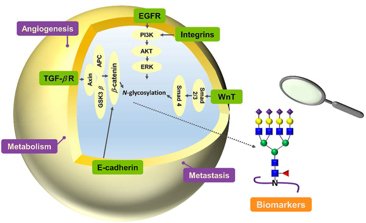
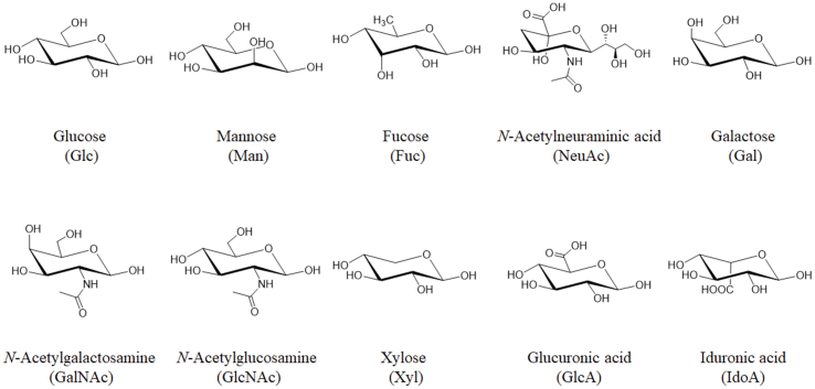
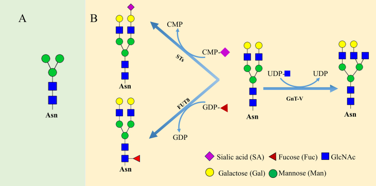
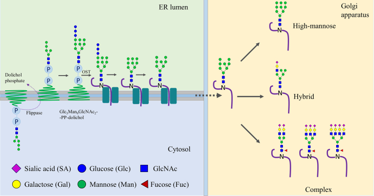
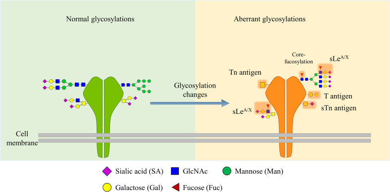
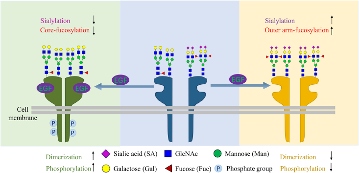
Despite advances in understanding the development and progression of cancer in recent years, there remains a lack of comprehensive characterization of the cancer glycoproteome. Glycoproteins play an important role in medicine and are involved in various human disease conditions including cancer. Glycan-moieties participate in fundamental cancer processes like cell signaling, invasion, angiogenesis, and metastasis. Aberrant N-glycosylation significantly impacts cancer processes and targeted therapies in clinic. Therefore, understanding N-glycosylation in a tumor is essential for comprehending disease progression and discovering anti-cancer targets and biomarkers for therapy monitoring and diagnosis. This review presents the fundamental process of protein N-glycosylation and summarizes glycosylation changes in tumor cells, including increased terminal sialylation, N-glycan branching, and core-fucosylation. Also, the role of N-glycosylation in tumor signaling pathways, migration, and metabolism are discussed. Glycoproteins and glycopeptides as potential biomarkers for early detection of cancer based on site specificity have been introduced. Collectively, understanding and exploring the cancer glycoproteome, along with its role in medicine, implication in cancer and other human diseases, highlights the significance of N-glycosylation in tumor processes, necessitating further research for potential anti-cancer targets and biomarkers.

Keywords: Cancer angiogenesis; Cancer biomarkers; Cancer metabolism; Cancer signaling; N-Glycosylated proteins.


Conflict of interest statement

The authors declare no conflicts of interest.

Figures

Image 1
Graphical abstract
Figure 1
Figure 1
Structures and names of 10 monosaccharides in humans which make up glycans.
Figure 2
Figure 2
(A) Five monosaccharides core structure GlcNAc2Man3. (B) Procedure of sialylation, core-fucosylation and branching of glycan chain are enzymatically catalyzed by sialyltransferases, α1,6-fucosyltransferase (FUT8) and N-acetylglucosaminyltransferase-V (GnT-V) by transferring the monosugar moieties from activated CMP-SA, GDP-Fuc and UDP-GlcNAc, respectively.
Figure 3
Figure 3
N-Glycosylation initiates in the ER and is elaborated in Golgi apparatus. The N-glycosylation is started by the transfer of GlcNAc2Man5 to dolichol phosphate on the cytosolic side of ER, and then flip to the luminal side by flippase. After adding four mannose and three glucose forming Glc3Man9GlcNAc2-PP-dolichol, the glycan moiety is then transferred to a sequon N-X-S-T of a nascent peptide chain by oligosaccharyltransferase (OST) complex. Next, three terminal glucose and one mannose are removed from the nascent glycopeptide, and the glycopeptide enters the Golgi apparatus to further trimming and branching, and thus produce the glycopeptide with diverse glycans.
Figure 4
Figure 4
Changes of glycosylation during tumor progression. During malignancy, aberrant glycosylation displays abnormal expression of truncated glycans such as T antigen, Tn and their sialylated forms ST and STn, respectively, as well as the multiple branching N-glycans with terminal sialylation.
Figure 5
Figure 5
Core-fucosylation promotes EGFR dimerization, whereas sialylation and outer-arm fucosylation suppress the EGFR dimerization when the ligand EGF binds to the EGFR.
Figure 6
Figure 6
Lectin recognition of glycosylation changes of tumor cells. Different lectin types expressed from cells are depicted, as well as the aberrant glycosylations in cancer cells. DC, dendritic cell; MGL, macrophage galactose-specific lectin; DC-SIGN, dendritic cell (DC)-specific ICAM-3-grabbing non-integrin.

Similar articles

Cited by

References

    1. Cancer . Feb 3, 2022. World health organization.https://www.who.int/news-room/fact-sheets/detail/cancer Available from:
    1. Cho K.C., Chen L., Hu Y., Schnaubelt M., Zhang H. Developing workflow for simultaneous analyses of phosphopeptides and glycopeptides. ACS Chem Biol. 2019;14:58–66. - PMC - PubMed
    1. Adamczyk B., Tharmalingam T., Rudd P.M. Glycans as cancer biomarkers. Biochim Biophys Acta Gen Subj. 2012;1820:1347–1353. - PubMed
    1. Büll C., den Brok M.H., Adema G.J. Sweet escape: sialic acids in tumor immune evasion. Biochim Biophys Acta Rev Cancer. 2014;1846:238–246. - PubMed
    1. Wu X., Zhao J., Ruan Y., Sun L., Xu C., Jiang H. Sialyltransferase ST3GAL1 promotes cell migration, invasion, and TGF-β1-induced EMT and confers paclitaxel resistance in ovarian cancer. Cell Death Dis. 2018;9:1102. - PMC - PubMed

LinkOut - more resources