Review
. 2024 Feb 29:15:1314507.
doi: 10.3389/fimmu.2024.1314507. eCollection 2024.

Longitudinal rheumatoid factor autoantibody responses after SARS-CoV-2 vaccination or infection

Affiliations
Review

Longitudinal rheumatoid factor autoantibody responses after SARS-CoV-2 vaccination or infection

Sofie Keijzer et al. Front Immunol. .

Abstract

Background: Rheumatoid factors (RFs) are autoantibodies that target the Fc region of IgG, and are found in patients with rheumatic diseases as well as in the healthy population. Many studies suggest that an immune trigger may (transiently) elicit RF responses. However, discrepancies between different studies make it difficult to determine if and to which degree RF reactivity can be triggered by vaccination or infection.

Objective: We quantitatively explored longitudinal RF responses after SARS-CoV-2 vaccination and infection in a well-defined, large cohort using a dual ELISA method that differentiates between true RF reactivity and background IgM reactivity. In addition, we reviewed existing literature on RF responses after vaccination and infection.

Methods: 151 healthy participants and 30 RA patients were included to measure IgM-RF reactivity before and after SARS-CoV-2 vaccinations by ELISA. Additionally, IgM-RF responses after a SARS-CoV-2 breakthrough infection were studied in 51 healthy participants.

Results: Published prevalence studies in subjects after infection report up to 85% IgM-RF seropositivity. However, seroconversion studies (both infection and vaccination) report much lower incidences of 2-33%, with a trend of lower percentages observed in larger studies. In the current study, SARS-CoV-2 vaccination triggered low-level IgM-RF responses in 5.5% (8/151) of cases, of which 1.5% (2/151) with a level above 10 AU/mL. Breakthrough infection was accompanied by development of an IgM-RF response in 2% (1/51) of cases.

Conclusion: Our study indicates that de novo RF induction following vaccination or infection is an uncommon event, which does not lead to RF epitope spreading.

Keywords: SARS-CoV-2; autoantibodies; autoimmunity; infection; rheumatoid arthritis; rheumatoid factor; vaccination.


Conflict of interest statement

TR and GW are inventors on a patent application based on the use of bioengineered IgG targets for the characterization of rheumatoid factor reactivity patterns. The remaining authors declare that the research was conducted in the absence of any commercial or financial relationships that could be construed as a potential conflict of interest.

Figures

Figure 1
Figure 1
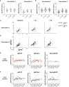
Longitudinal IgM-RF responses after SARS-CoV-2 vaccination. (A) Schematic overview of serum sample collection after vaccination. Timepoint V1 + 10d and V2 + 10d were collected for a subset of the healthy participant group. Median interval time for healthy participants between V1 and V2 is 42 days (IQR: 36-42), and between V2 and V3 190 days (IQR: 181-200). For RA patients median interval time between V1 and V2 is 37 days (IQR: 35-42), and between V2 and V3 185 days (IQR: 116-201). (B) Study participant baseline characteristics in the SARS-CoV-2 vaccination cohort and breakthrough infection cohort. RA patients were treated with immunosuppressive medication; 21/30 (70%) used methotrexate, 3/30 (10%) methotrexate + TNF inhibitors, and 6/30 (20%) other immunosuppressive medication. (C) IgM-RF levels (left panel) and IgM-RF levels against IgG-Bare (right panel) at different timepoints after vaccination in healthy participants. No difference in time points was observed based on Kruskal-Wallis test on the frequencies of positive samples per time point. (D) IgM-RF levels (left panel) and IgM-RF levels against IgG-Bare (right panel) at different timepoints after vaccination in RA patients. The median IgM-RF level for each timepoint is indicated in red. No significant differences in IgM-RF levels were observed between timepoints (Paired one-way ANOVA; F (2.108, 60.08) = 0.5783; P = 0.57) (E) IgM-RF reactivity against WT IgG and IgG-Bare, resembling the true and background target binding of IgM-RF, after vaccination in healthy participants and RA patients. Individuals with an IgM-RF level above the cutoff (>3 AU/mL) at baseline are indicated in grey to make tracking of individuals over time easier. ns, not significant.
Figure 2
Figure 2
Early IgM-RF responses after SARS-CoV-2 vaccination. (A) IgM-RF levels (left panel) and IgM-RF levels against IgG-Bare (right panel) at 10 days and 28 days after the first SARS-CoV-2 vaccination in a subset of healthy participants (n=45). (B) IgM-RF levels (left panel) and IgM-RF levels against IgG-Bare (right panel) at 10 days and 28 days after the second SARS-CoV-2 vaccination in a subset of healthy participants (n=80). Frequencies of positive samples per time point were analyzed using Kruskal-Wallis test, * < 0.05. (C, D) IgM-RF reactivity against WT IgG and IgG-Bare, resembling the true and background target binding of IgM-RF, 10 days and 28 days after first vaccination (panel C) and second vaccination (panel D) in healthy participants. Individuals with an IgM-RF level above the cutoff (>3 AU/mL) at baseline are indicated in grey to make tracking of individuals over time easier. (E, F) Cases of vaccine-induced IgM-RF responses. We selected individuals in which WT IgM-RF levels increased >2-fold relative to baseline, thereby reaching levels above 3 AU/mL, at any time point after vaccination. Of these 15 cases, seven also developed background IgM-Bare reactivity, suggesting the increase in IgM-RF reactivity does not represent true RF (‘false positive’; panel E), whereas eight cases developed only WT IgM-RF reactivity, meaning a true positive increase in IgM-RF reactivity (panel F). IgM-RF levels (left panel), IgM-RF levels against IgG-Bare (middle panel) and delta of IgM-RF minus background IgM-Bare levels (right panel) at different timepoints are shown. For the delta IgM-RF levels, extremely low and negative delta levels are set at 1 AU/ml.
Figure 3
Figure 3
IgM-RF responses after SARS-CoV-2 breakthrough infection. (A) Schematic overview of serum sample collection after breakthrough infection. (B) IgM-RF levels (left panel) and IgM-RF levels against IgG-Bare (right panel) at different timepoints after infection in healthy participants. (C) IgM-RF and IgM-Bare reactivity after breakthrough infection. Individuals with an IgM-RF level above the cutoff (>3 AU/mL) at baseline are indicated in grey to make tracking of individuals over time easier.

References

    1. Newkirk MM. Rheumatoid factors: Host resistance or autoimmunity? Clin Immunol. (2002) 104:1–13. doi: 10.1006/clim.2002.5210 - DOI - PubMed
    1. Van Schaardenburg D, Lagaay AM, Otten HG, Breedveld FC. The relation between class-specific serum rheumatoid factors and age in the general population. Br J Rheumatol. (1993) 32:546–9. doi: 10.1093/rheumatology/32.7.546 - DOI - PubMed
    1. Sobhy N, Ghoniem SA, Eissa BM, Kamal A, Medhat A, Elsaid NY. Disease characteristics in high versus low titers of rheumatoid factor or anti-citrullinated peptide antibody in rheumatoid arthritis patients. Egypt Rheumatol. (2022) 44:325–8. doi: 10.1016/j.ejr.2022.04.004 - DOI
    1. Van Snick JL, Van Roost E, Markowetz B, Cambiaso CL, Masson PL. Enhancement by IgM rheumatoid factor of in vitro ingestion by macrophages and in vivo clearance of aggregated IgG or antigen-antibody complexes. Eur J Immunol. (1978) 8:279–85. doi: 10.1002/eji.1830080412 - DOI - PubMed
    1. Hogben DN, Devey ME. Studies on rheumatoid factor: I. The effect of rheumatoid factor on the clearance of preformed immune complexes in mice. Clin Exp Immunol. (1986) 66:648–53. - PMC - PubMed

Publication types