

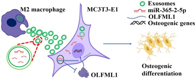
Macrophage exosomes modified by miR-365-2-5p promoted osteoblast osteogenic differentiation by targeting OLFML1
- PMID: 38487712
- PMCID: PMC10939467
- DOI: 10.1093/rb/rbae018
Macrophage exosomes modified by miR-365-2-5p promoted osteoblast osteogenic differentiation by targeting OLFML1
Abstract
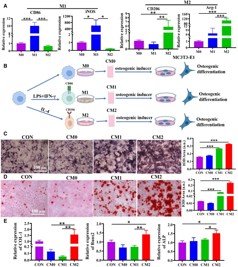
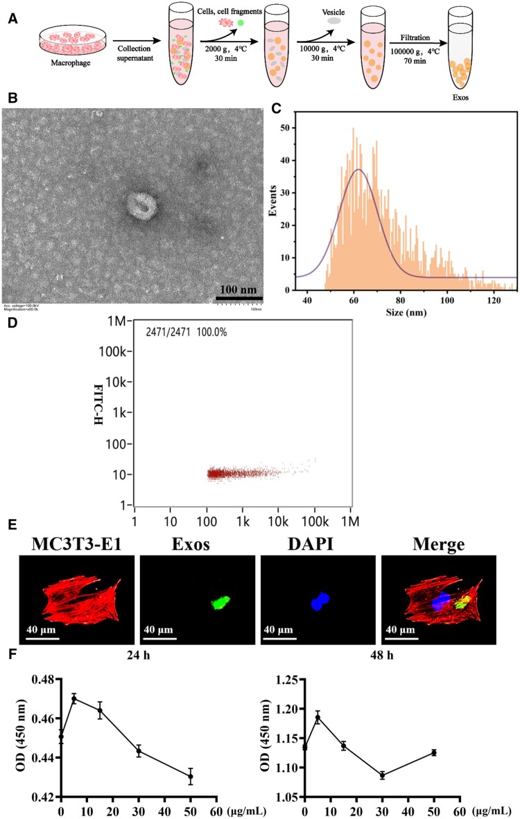
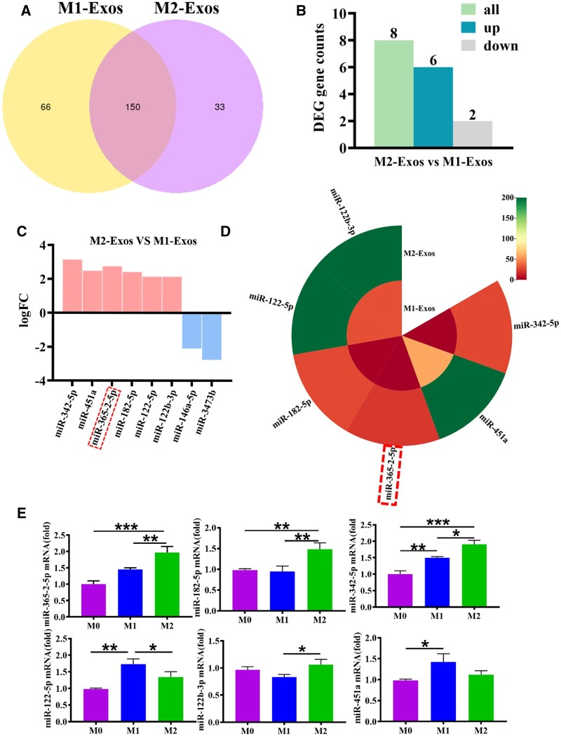
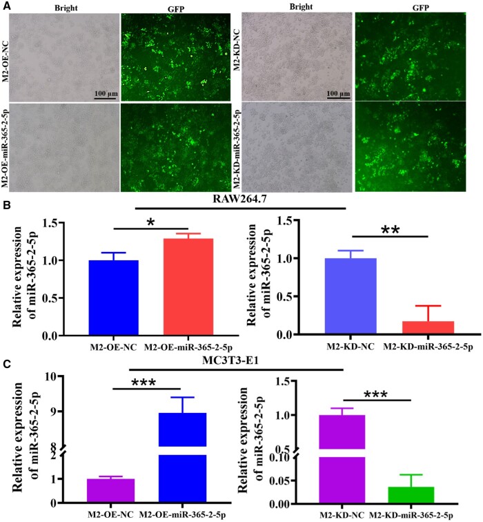
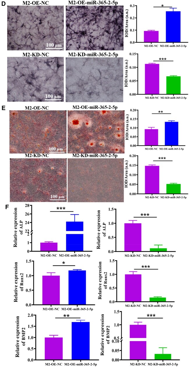
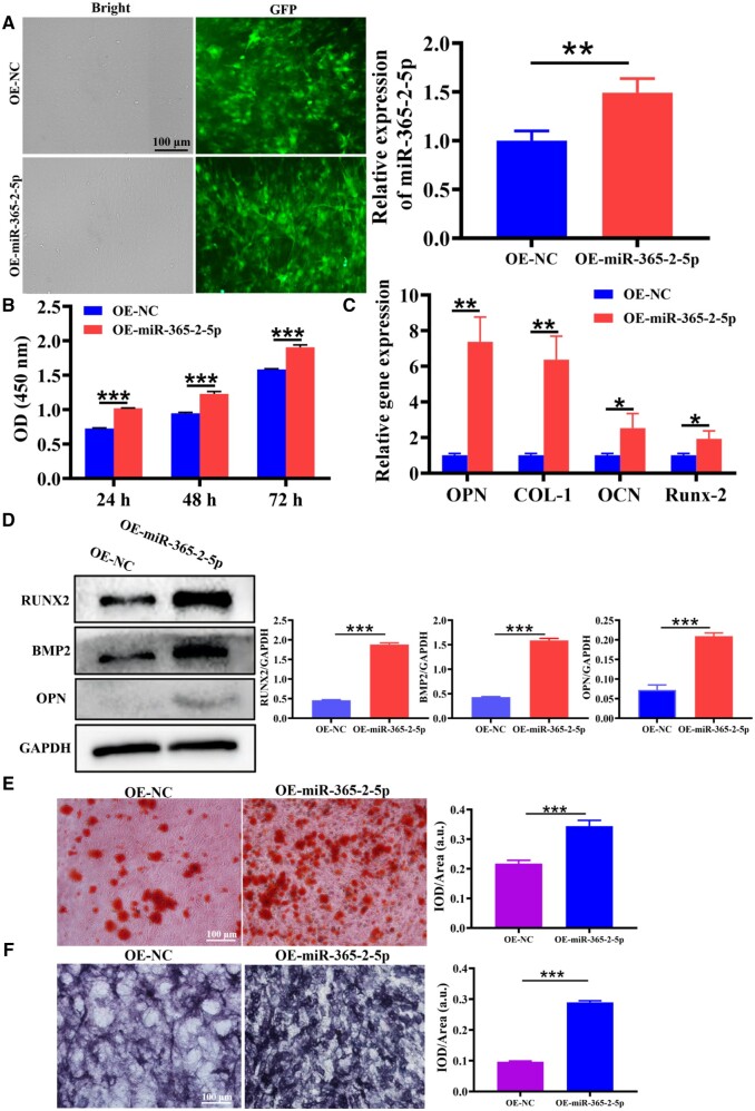
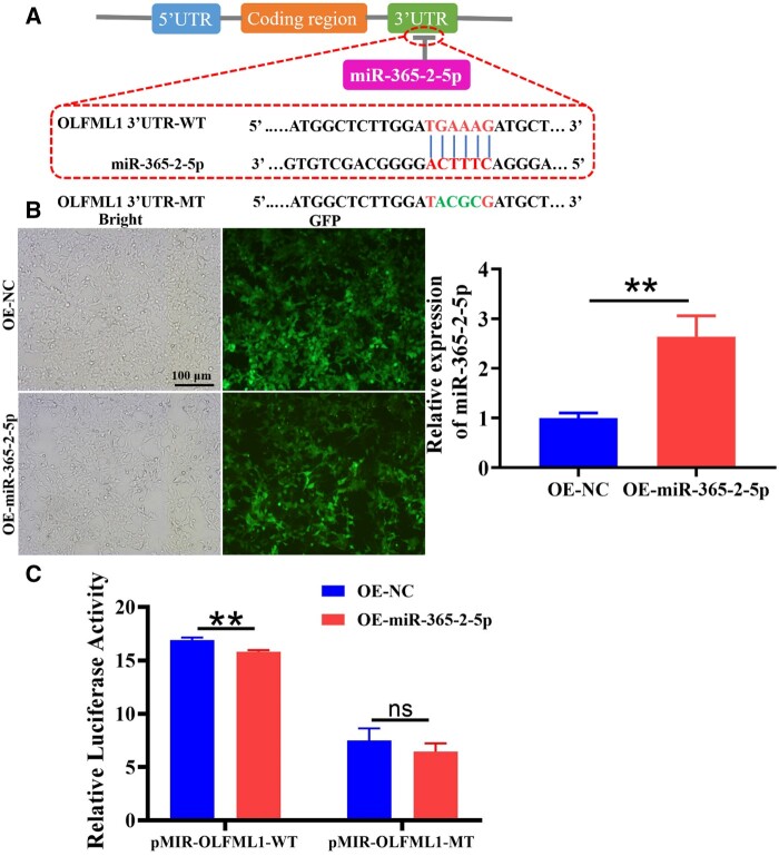
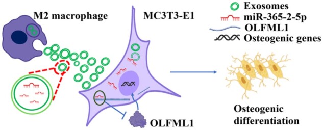
In the bone immune microenvironment, immune cells can regulate osteoblasts through a complex communication network. Macrophages play a central role in mediating immune osteogenesis, exosomes derived from them have osteogenic regulation and can be used as carriers in bone tissue engineering. However, there are problems with exosomal therapy alone, such as poor targeting, and the content of loaded molecules cannot reach the therapeutic concentration. In this study, macrophage-derived exosomes modified with miR-365-2-5p were developed to accelerate bone healing. MC3T3-E1 cells were incubated with the culture supernatants of M0, M1 and M2 macrophages, and it was found that the culture medium of M2 macrophages had the most significant effects in contributing to osteogenesis. High-throughput sequencing identified that miR-365-2-5p was significantly expressed in exosomes derived from M2 macrophages. We incubated MC3T3-E1 with exosomes overexpressing or knocking down miR-365-2-5p to examine the biological function of exosome miR-365-2-5p on MC3T3-E1 differentiation. These findings suggested that miR-365-2-5p secreted by exosomes increased the osteogenesis of MC3T3-E1. Moreover, miR-365-2-5p had a direct influence over osteogenesis for MC3T3-E1. Sequencing analysis combined with dual luciferase detection indicated that miR-365-2-5p binded to the 3'-UTR of OLFML1. In summary, exosomes secreted by M2 macrophages targeted OLFML1 through miR-365-2-5p to facilitate osteogenesis.
Keywords: exosomes; macrophage; osteogenesis; osteoimmunology.
© The Author(s) 2024. Published by Oxford University Press.
Figures
Similar articles
-
MicroRNA-21a-5p-modified macrophage exosomes as natural nanocarriers promote bone regeneration by targeting GATA2.Regen Biomater. 2023 Aug 31;10:rbad075. doi: 10.1093/rb/rbad075. eCollection 2023. Regen Biomater. 2023. PMID: 37719929 Free PMC article.
-
Involvement of MiRNA-211-5p and Arhgap11a Interaction During Osteogenic Differentiation of MC3T3-E1 Cells.Front Surg. 2022 Apr 15;9:857170. doi: 10.3389/fsurg.2022.857170. eCollection 2022. Front Surg. 2022. PMID: 35495761 Free PMC article.
-
Hyperin up-regulates miR-7031-5P to promote osteogenic differentiation of MC3T3-E1 cells.Histol Histopathol. 2023 Oct;38(10):1219-1229. doi: 10.14670/HH-18-579. Epub 2022 Dec 30. Histol Histopathol. 2023. PMID: 36633331
-
M2 macrophage-derived exosomal miR-486-5p influences the differentiation potential of bone marrow mesenchymal stem cells and osteoporosis.Aging (Albany NY). 2023 Sep 25;15(18):9499-9520. doi: 10.18632/aging.205031. Epub 2023 Sep 25. Aging (Albany NY). 2023. PMID: 37751585 Free PMC article.
-
Comprehensive analysis of M2 macrophage-derived exosomes facilitating osteogenic differentiation of human periodontal ligament stem cells.BMC Oral Health. 2022 Dec 27;22(1):647. doi: 10.1186/s12903-022-02682-5. BMC Oral Health. 2022. PMID: 36575449 Free PMC article.
Cited by
-
Stabilization of OLFML1 via m6A Reader IGF2BP3 Drives CSC Characteristics Through Hedgehog Pathway Activation in CRC.Int J Biol Sci. 2025 Jun 23;21(10):4334-4352. doi: 10.7150/ijbs.111032. eCollection 2025. Int J Biol Sci. 2025. PMID: 40765816 Free PMC article.
-
Strontium-incorporated hydroxyapatite nanocomposites promoting bone formation and angiogenesis by modulating M2 macrophage polarization in the bone microenvironment.Regen Biomater. 2025 Jun 23;12:rbaf066. doi: 10.1093/rb/rbaf066. eCollection 2025. Regen Biomater. 2025. PMID: 40799901 Free PMC article.
-
Exosome Source Matters: A Comprehensive Review from the Perspective of Diverse Cellular Origins.Pharmaceutics. 2025 Jan 22;17(2):147. doi: 10.3390/pharmaceutics17020147. Pharmaceutics. 2025. PMID: 40006514 Free PMC article. Review.
-
M2 macrophage-derived exosomes enable osteogenic differentiation and inhibit inflammation in human periodontal ligament stem cells through promotion of CXCL12 expression.BMC Oral Health. 2024 Sep 11;24(1):1070. doi: 10.1186/s12903-024-04831-4. BMC Oral Health. 2024. PMID: 39261847 Free PMC article.
-
Fused exosomal targeted therapy in periprosthetic osteolysis through regulation of bone metabolic homeostasis.Bioact Mater. 2025 Apr 8;50:171-188. doi: 10.1016/j.bioactmat.2025.04.006. eCollection 2025 Aug. Bioact Mater. 2025. PMID: 40248188 Free PMC article.
References
-
- Huang EE, Zhang N, Ganio EA, Shen H, Li X, Ueno M, Utsunomiya T, Maruyama M, Gao Q, Su N, Yao Z, Yang F, Gaudillière B, Goodman SB.. Differential dynamics of bone graft transplantation and mesenchymal stem cell therapy during bone defect healing in a murine critical size defect. J Orthop Translat 2022;36:64–74. - PMC - PubMed
-
- Wu P, Shen L, Liu HF, Zou XH, Zhao J, Huang Y, Zhu YF, Li ZY, Xu C, Luo LH, Luo ZQ, Wu MH, Cai L, Li XK, Wang ZG.. The marriage of immunomodulatory, angiogenic, and osteogenic capabilities in a piezoelectric hydrogel tissue engineering scaffold for military medicine. Mil Med Res 2023;10:35. - PMC - PubMed
-
- Louiselle AE, Niemiec SM, Zgheib C, Liechty KW.. Macrophage polarization and diabetic wound healing. Transl Res 2021;236:109–16. - PubMed
LinkOut - more resources
Full Text Sources
