. 2024 Jan 11;134(6):e172095.
doi: 10.1172/JCI172095.

TGF-β controls alveolar type 1 epithelial cell plasticity and alveolar matrisome gene transcription in mice

Affiliations

TGF-β controls alveolar type 1 epithelial cell plasticity and alveolar matrisome gene transcription in mice

Danielle A Callaway et al. J Clin Invest. .

Abstract

Premature birth disrupts normal lung development and places infants at risk for bronchopulmonary dysplasia (BPD), a disease disrupting lung health throughout the life of an individual and that is increasing in incidence. The TGF-β superfamily has been implicated in BPD pathogenesis, however, what cell lineage it impacts remains unclear. We show that TGFbr2 is critical for alveolar epithelial (AT1) cell fate maintenance and function. Loss of TGFbr2 in AT1 cells during late lung development leads to AT1-AT2 cell reprogramming and altered pulmonary architecture, which persists into adulthood. Restriction of fetal lung stretch and associated AT1 cell spreading through a model of oligohydramnios enhances AT1-AT2 reprogramming. Transcriptomic and proteomic analyses reveal the necessity of TGFbr2 expression in AT1 cells for extracellular matrix production. Moreover, TGF-β signaling regulates integrin transcription to alter AT1 cell morphology, which further impacts ECM expression through changes in mechanotransduction. These data reveal the cell intrinsic necessity of TGF-β signaling in maintaining AT1 cell fate and reveal this cell lineage as a major orchestrator of the alveolar matrisome.

Keywords: Extracellular matrix; Integrins; Pulmonology; Respiration.


Conflict of interest statement

Conflict of interest: The authors have declared that no conflict of interest exists.

Figures

Figure 1
Figure 1. TGF-β is involved in regulating AT1 cell fate during prenatal and postnatal late lung development.
(A) Tamoxifen was delivered to control heterozygous (TGFbr2fl/+) littermates and KO (TGFbr2fl/fl or TGFbr2AT1–KO) mice through IP injection of the pregnant dam at E15.5 and lungs were harvested at E18.5. (B) IHC for EYFP, HOPX, and SFTPC demonstrate increased AT1 reprogramming into AT2 cells after prenatal loss of TGFbr2. The yellow dashed box denotes the magnified region shown below the image and separated by fluorescence channel. (C) Quantification of lineage tracing in B denoting percent of cells that were EYFP+ and SFTPC+ by an unpaired 2-tailed t test (n = 5 per group). (D) IHC for NKX2.1, HOPX, and DCLAMP demonstrate no significant change in the AT1 cell composition after prenatal loss of TGFbr2. The yellow dashed box denotes the magnified region shown below the image and separated by fluorescence channel. (E) Quantification of AT2 cell numbers from total NKX2.1+ cells in (D) denoting percent of cells that were NKX2.1+ and DCLAMP+ and (F) the AT2/AT1 cell ratio at E18.5 by an unpaired 2-tailed t test (n = 6 per group). (G) In postnatal lineage-tracing experiments, control (HopxcreERT2:R26REYFP) and TGFbr2AT1–KO newborn pups (P0) were injected with tamoxifen and the lungs were harvested at P5 and P42. (H) IHC for EYFP, HOPX, and SFTPC demonstrate increased AT1 reprogramming into AT2 cells after prenatal loss of TGFbr2 at both P5 (top) and P42 (bottom). The yellow dashed box denotes the magnified region shown below the image and separated by fluorescence channel. (I) Quantification of lineage tracing in H denoting percent of cells that were EYFP+ and SFTPC+ at P5 (left) and P42 (right) by unpaired 2-tailed t tests with Welch’s correction (n = 7–12 per group). Each dot represents a single mouse. Scale bars: 10 μm. P values are denoted above the plots. Schematics in A and G were created in BioRender.
Figure 2
Figure 2. TGF-β is involved in maintaining normal pulmonary architecture during late lung development.
(A) H&E images of lungs from E18.5 mice following tamoxifen injection at E15.5 show that TGFbr2AT1–KO embryonic lungs compared with heterozygous littermates demonstrate increased mean septal thickness. Red-dashed boxes indicate the zoomed area shown in the top-right of the image. (B) Quantification of mean septal thickness (μm) from A by an unpaired 2-tailed t test (n = 5–10 per group). (C) H&E images of lungs from P5 (top) and P42 (bottom) mice following tamoxifen injection at P0 indicate that loss of TGF-β postnatally results in alveolar simplification and increased mean septal thickness. Red-dashed boxes indicate the zoomed area shown in the top-right of the image. (D) Quantification of MLI (μm) and (E) mean septal thickness (μm) by unpaired 2-tailed t tests with Welch’s correction (n = 8–10 per group). Each dot represents a single mouse. Scale bars: 50 μm. P values are denoted above the plots.
Figure 3
Figure 3. Oligohydramnios alters AT1 cell numbers through increased AT1-AT2 cell reprogramming.
(A) Oligohydramnios was induced through amniotic fluid reduction at E15.5 followed by injection of tamoxifen to enable lineage tracing. Lungs from the Control or AT1-KO fetal mice were harvested at E18.5 for further analysis. (B) Amniotic fluid (green) was aspirated from amniotic sacs in the right uterine horn. (C) IHC for lineage-tracing with EYFP, HOPX, and SFTPC demonstrate that there is increased AT1-AT2 cell reprogramming with oligohydramnios although not to a significant extent in AT1-KO pups. The yellow dashed box denotes the magnified region shown below the image and separated by fluorescence channel. (D) Quantification of lineage tracing in C denoting percent of cells that were EYFP+ and SFTPC+ by a 2-way ANOVA with Tukey’s post test for multiple comparisons (n = 4–5 per group). (E) H&E images of control (left) and AT1-KO (right) who underwent oligohydramnios (bottom) or were the littermate controls (top) lungs reveal that lack of amniotic fluid leads to increased mean septal thickness. (F) Quantification of mean septal thickness (μm) by a 2-way ANOVA with Tukey’s post test for multiple comparisons (n = 4–5 per group). Each dot represents a single mouse. Scale bars: 50 μm for H&E and 10 μm for IHC. P values are denoted above the plots. Schematics in A and B were created in BioRender.
Figure 4
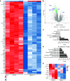
Figure 4. Transcriptomic and proteomic profiling reveal a role for TGFβ in regulating AT1 cell expression of the pulmonary matrisome.
(A) Heatmap of differentially expressed genes (upregulated on top, downregulated on bottom) from AT1 cells of control heterozygous littermates (left) or TGFbr2AT1–KO (right) mice at P5 (n = 4). Genes depicted were filtered with a cut off of 0.05 for the P value and log FC of 2. (B) Volcano plot of differentially expressed genes from RNA-Seq from (A) with ECM-related genes labeled reveal that several are significantly downregulated with loss of TGFbr2. (C) GO enrichment for cellular component and (D) molecular function of downregulated genes reveal several ECM-related terms. (E) Heatmap of ECM-related proteins from proteomics results of control heterozygous littermates (left) or TGFbr2AT1–KO (right) AT1 cells obtained at P5 (n = 3).
Figure 5
Figure 5. AT1 cells express collagen 4 in a time-dependent manner and are important hubs for communication across the developing lung.
(A) UMAP of scRNA-Seq data at P3 from Zepp et al, 2021 (40) of collagen IV subtype expression across the mouse lung including Col4a1, (B) Col4a3, and (C) Col4a4, denoting that the latter are highly specific for AT1 cells. (D) Evaluation of previously generated scRNA-Seq data across development indicates that AT1-enriched collagen IV and proteoglycan genes become highly expressed in the postnatal period through adulthood indicating a role for AT1 cells in the active production and remodeling of the pulmonary matrisome across the lifespan. (E) Circle plot from CellChat analysis of outgoing communication from AT1 and AT2 cells at P3 (from Zepp et al, 2021 data set) (40), to cells of the mesenchyme (ASM_SCMF, Wnt2-pa, VSM, AMP, and MANCs) and to the endothelium (CapEC), denoting that AT1 cells are the primary communicators of the alveolar epithelium.
Figure 6
Figure 6. Loss of TGF-β at birth perturbs AT1-mediated matrisome expression through adulthood.
(A) RNA FISH for Col4a3 and Hopx with IHC for lineage-labeled cells at P5 (top) and P42 (bottom). The yellow dashed box denotes the magnified region shown below the image and separated by fluorescence channel. (B) TGFbr2AT1–KO AT1 cells exhibit decreased qPCR RNA (FC compared with GAPDH, normalized to controls) transcript expression for several AT1 cell-enriched core matrisome constituents including collagens Col4a3 and (D) Col4a4 and glycoproteins (F) Lamb3 and (H) Igfbp7 at P5 that persists to P42 (n = 5–7, unpaired 2-tailed t test with Welch’s correction). (C) RNA FISH of P5 lungs for Col4a4 with IHC for lineage-labeled cells. The yellow dashed box denotes the magnified region shown below the image and separated by fluorescence channel. (E) RNA FISH for Hopx and glycoproteins Lamb3 and (G) Igfbp7 with IHC for lineage-labeled cells. The yellow dashed box denotes the magnified region shown below the image and separated by fluorescence channel. Each dot represents a single mouse. Scale bars: 10 μm. P values are denoted above the plots.
Figure 7
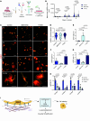
Figure 7. TGF-β–mediated integrin binding regulates AT1 cell size, morphology, and ECM expression.
(A) To obtain AT1 cells for culture, P5–P10 pup lungs were obtained after lineage labeling with tamoxifen at P0. Whole lung cell suspensions were obtained using a dispase, DNase, and collagenase digestion buffer after which the epithelial cell population was enriched by depleting the CD45+ and CD31+ population. Remaining cells were fluorescently sorted with FACS to obtain a CD326+ and YFP+ suspension. Cells were plated onto fibronectin-coated plates with or without TGF-β1 ligand (7.5 ng/mL) or TGF-β inhibitor SB431542 (10μM) and evaluated at days 2, 4, and 6. (B) ICC of RAGE+ (AGER+) cells treated with TGF-β ligand or inhibitor in culture at days 2, 4, and 6 with a zoomed image of cells at day 6 appearing at the bottom. Scale bars: 100 μm. (C) Quantification of mean AGER+ cell area depicted in B by 1-way ANOVA with Holms Šidák’s test for multiple comparisons (n = 135–231). (D) Quantification of mean cell roundness at day 6 depicted in B by 1-way ANOVA with Holms Šidák’s test for multiple comparisons (n = 148–167). (E) Quantification of qPCR RNA transcript expression levels (FC compared with GAPDH, normalized to controls) of the AT2 marker Sftpb, fibronectin-binding integrins (F) Itga5 and (G) Itgb1, and (H) basement membrane constituents including the collagen IV subtypes Col4a1, Col4a3, and Col4a4 and laminin-332 constituents Lama3 and Lamb3 (n = 3 per group, 1-way ANOVA with Tukey’s multiple comparisons). (I) Representative schematic of findings indicating that TGF-β regulates integrin expression to guide ECM binding and cellular spread, which affects cell identity and matrisome expression and impacts lung development. Schematics in A and I were created in BioRender. Results are representative of 3 experiments.

Update of

References

    1. Thébaud B, et al. Bronchopulmonary dysplasia. Nat Rev Dis Primers. 2019;5(1):78. doi: 10.1038/s41572-019-0127-7. - DOI - PMC - PubMed
    1. Bell EF, et al. Mortality, in-hospital morbidity, care practices, and 2-year outcomes for extremely preterm infants in the US, 2013-2018. JAMA. 2022;327(3):248–263. doi: 10.1001/jama.2021.23580. - DOI - PMC - PubMed
    1. Resch B, et al. Prematurity and the burden of influenza and respiratory syncytial virus disease. World J Pediatr. 2016;12(1):8–18. doi: 10.1007/s12519-015-0055-x. - DOI - PubMed
    1. Homaira N, et al. Impact of influenza on hospitalization rates in children with a range of chronic lung diseases. Influenza Other Respir Viruses. 2019;13(3):233–239. doi: 10.1111/irv.12633. - DOI - PMC - PubMed
    1. vom Hove M, et al. Pulmonary outcome in former preterm, very low birth weight children with bronchopulmonary dysplasia: a case-control follow-up at school age. J Pediatr. 2014;164(1):40–45. doi: 10.1016/j.jpeds.2013.07.045. - DOI - PubMed

MeSH terms

Substances