. 2024 Jun;35(2):113-121.
doi: 10.1007/s00335-024-10033-8. Epub 2024 Mar 15.

Generation of a humanized mAce2 and a conditional hACE2 mouse models permissive to SARS-COV-2 infection

Affiliations

Generation of a humanized mAce2 and a conditional hACE2 mouse models permissive to SARS-COV-2 infection

I-Wen Song et al. Mamm Genome. 2024 Jun.

Abstract

The Severe Acute Respiratory Syndrome Coronavirus-2 (SARS-CoV-2) remains a public health concern and a subject of active research effort. Development of pre-clinical animal models is critical to study viral-host interaction, tissue tropism, disease mechanisms, therapeutic approaches, and long-term sequelae of infection. Here, we report two mouse models for studying SARS-CoV-2: A knock-in mAce2F83Y,H353K mouse that expresses a mouse-human hybrid form of the angiotensin-converting enzyme 2 (ACE2) receptor under the endogenous mouse Ace2 promoter, and a Rosa26 conditional knock-in mouse carrying the human ACE2 allele (Rosa26hACE2). Although the mAce2F83Y,H353K mice were susceptible to intranasal inoculation with SARS-CoV-2, they did not show gross phenotypic abnormalities. Next, we generated a Rosa26hACE2;CMV-Cre mouse line that ubiquitously expresses the human ACE2 receptor. By day 3 post infection with SARS-CoV-2, Rosa26hACE2;CMV-Cre mice showed significant weight loss, a variable degree of alveolar wall thickening and reduced survival rates. Viral load measurements confirmed inoculation in lung and brain tissues of infected Rosa26hACE2;CMV-Cre mice. The phenotypic spectrum displayed by our different mouse models translates to the broad range of clinical symptoms seen in the human patients and can serve as a resource for the community to model and explore both treatment strategies and long-term consequences of SARS-CoV-2 infection.


Conflict of interest statement

Competing Interests Declaration: The authors have no competing interests to disclose.

Figures

Figure 1.
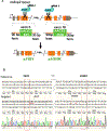
Figure 1.. Generation of the mouse-human hybrid mAce2F83Y,H353K mouse model.
(A) Schematic presentation of the approach to generate a mouse-human hybrid allele via CRISPR/Cas9 gene editing. In brief, two guide RNAs directed Cas9-mediated cleavage at codon 83 (exon 3) and codon 353 (exon 18) of Ace2. Edits were introduced by two single-stranded donor DNA oligos. (B) Sanger sequencing confirmed correct editing of the mAce2 allele in a male founder (red boxes). Of note, knocked-in mutations are in obligate cis configuration (14 kb apart) since mAce2 is located on the X chromosome. Silent mutations introducing restriction enzyme sites for genotyping are underlined in red. (C) Expression of Ace2 in multiple tissues, as measured by real-time qPCR in mAce2F83Y,H353K mice and wild type littermates (WT) and normalized to beta actin, showing that gene editing did not significantly alter the expression level. The ratio of mAce2F83Y,H353K to endogenous (WT) mAce2 level is plotted in various tissues. Data shown as mean +/− SD, n=3–4 per tissue.
Figure 2.
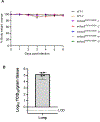
Figure 2.. SARS-CoV-2 infection in mAce2F83Y,H353K mouse model.
(A) Weight has not changed significantly over 6 days post infection. Data presented as proportion of starting weight (weight at day 0). (B) Measurement of viral load in mutant mice lungs indicated successful inoculation in 4/5 mice (measurement was below limit of detection in 1/5 mice). Data presented as viral titer per gram lung tissue. Limit of detection (LOD) for tissue viral titer is Log10 1.69 TCID50/gm.
Figure 3.
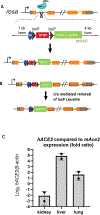
Figure 3.. Targeting the hACE2 cDNA to the Rosa26 locus to generate a conditional Rosa26hACE2 mouse model.
(A) Schematic presentation of the approach to generate the targeted Rosa26 allele via CRISPR/Cas9 gene editing. Human ACE2 cDNA was cloned into the Rosa26-DEST vector downstream of a loxP-polyA-loxP stop cassette. (B) Expression of the hACE2 allele will be activated upon Cre-mediated removal of the loxP cassette. (C) Expression of hACE2 in kidney, liver and lung tissues, as measured by real-time qPCR in Rosa26hACE2;CMV-Cre mice. Graph presents the ratio of hACE2 to endogenous (WT) mAce2 level in various tissues. Data shown as mean +/− SD, n=2 per tissue.
Figure 4.
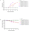
Figure 4.. SARS-CoV-2 infection leads to significant morbidity and mortality in Rosa26hACE2;CMV-Cre mouse model.
Outcome of viral infection was assessed daily up to 6 days post infection, using (A) Clinical scoring system, ranging in severity from 1 (well-appearing) to 4 (severely affected), (B) Change in body weight post infection, presented as proportion of starting weight (weight at day 0).; and (C) Kaplan-Meier survival curve, demonstrating morbidity and lethality in 4 out of 5 Rosa26hACE2;CMV-Cre mice by day 4–6 post infection. (D) Viral titers measured in lung and brain tissue from Rosa26hACE2;CMV-Cre mice on day 3 post infection (data presented as viral titer per gram lung tissue, n= 4). Limit of detection (LOD) for tissue viral titer is Log10 1.69 TCID50/gm. (E) Hematoxylin-stained histological lung sections of SARS-CoV-2 treated Rosa26hACE2;CMV-Cre mice on day 3 post infection (x20, Scale bar 200uM). Bottom panel: Magnification of the insert area (marked in red), showing mild alveolar wall thickening (red arrows) in the mutant mice.

References

    1. Akkiz H (2023) Unraveling the Molecular and Cellular Pathogenesis of COVID-19-Associated Liver Injury. Viruses 15 - PMC - PubMed
    1. Al-Aly Z, Xie Y, Bowe B (2021) High-dimensional characterization of post-acute sequelae of COVID-19. Nature 594, 259–264 - PubMed
    1. Bao L, Deng W, Huang B, Gao H, Liu J, Ren L, Wei Q, Yu P, Xu Y, Qi F, Qu Y, Li F, Lv Q, Wang W, Xue J, Gong S, Liu M, Wang G, Wang S, Song Z, Zhao L, Liu P, Zhao L, Ye F, Wang H, Zhou W, Zhu N, Zhen W, Yu H, Zhang X, Guo L, Chen L, Wang C, Wang Y, Wang X, Xiao Y, Sun Q, Liu H, Zhu F, Ma C, Yan L, Yang M, Han J, Xu W, Tan W, Peng X, Jin Q, Wu G, Qin C (2020) The pathogenicity of SARS-CoV-2 in hACE2 transgenic mice. Nature 583, 830–833 - PubMed
    1. Brielle ES, Schneidman-Duhovny D, Linial M (2020) The SARS-CoV-2 Exerts a Distinctive Strategy for Interacting with the ACE2 Human Receptor. Viruses 12 - PMC - PubMed
    1. Carofino BL, Justice MJ (2015) Tissue-Specific Regulation of Oncogene Expression Using Cre-Inducible ROSA26 Knock-In Transgenic Mice. Curr Protoc Mouse Biol 5, 187–204 - PMC - PubMed

Substances

LinkOut - more resources