

Recommender-based bone tumour classification with radiographs-a link to the past
- PMID: 38488971
- PMCID: PMC11399296
- DOI: 10.1007/s00330-024-10672-0
Recommender-based bone tumour classification with radiographs-a link to the past
Abstract
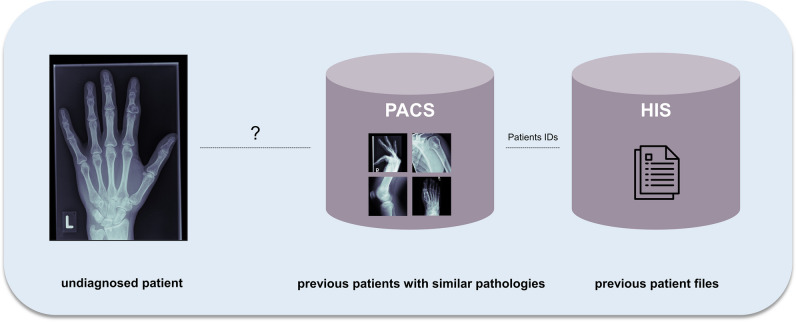
Objectives: To develop an algorithm to link undiagnosed patients to previous patient histories based on radiographs, and simultaneous classification of multiple bone tumours to enable early and specific diagnosis.
Materials and methods: For this retrospective study, data from 2000 to 2021 were curated from our database by two orthopaedic surgeons, a radiologist and a data scientist. Patients with complete clinical and pre-therapy radiographic data were eligible. To ensure feasibility, the ten most frequent primary tumour entities, confirmed histologically or by tumour board decision, were included. We implemented a ResNet and transformer model to establish baseline results. Our method extracts image features using deep learning and then clusters the k most similar images to the target image using a hash-based nearest-neighbour recommender approach that performs simultaneous classification by majority voting. The results were evaluated with precision-at-k, accuracy, precision and recall. Discrete parameters were described by incidence and percentage ratios. For continuous parameters, based on a normality test, respective statistical measures were calculated.
Results: Included were data from 809 patients (1792 radiographs; mean age 33.73 ± 18.65, range 3-89 years; 443 men), with Osteochondroma (28.31%) and Ewing sarcoma (1.11%) as the most and least common entities, respectively. The dataset was split into training (80%) and test subsets (20%). For k = 3, our model achieved the highest mean accuracy, precision and recall (92.86%, 92.86% and 34.08%), significantly outperforming state-of-the-art models (54.10%, 55.57%, 19.85% and 62.80%, 61.33%, 23.05%).
Conclusion: Our novel approach surpasses current models in tumour classification and links to past patient data, leveraging expert insights.
Clinical relevance statement: The proposed algorithm could serve as a vital support tool for clinicians and general practitioners with limited experience in bone tumour classification by identifying similar cases and classifying bone tumour entities.
Key points: • Addressed accurate bone tumour classification using radiographic features. • Model achieved 92.86%, 92.86% and 34.08% mean accuracy, precision and recall, respectively, significantly surpassing state-of-the-art models. • Enhanced diagnosis by integrating prior expert patient assessments.
Keywords: Bone neoplasms; Classification; Deep learning; Machine learning; Radiography.
© 2024. The Author(s).
Conflict of interest statement
The authors of this manuscript declare no relationships with any companies, whose products or services may be related to the subject matter of the article.
Figures


References
-
- Picci P, Manfrini M, Donati DM et al (2020) Diagnosis of musculoskeletal tumors and tumor-like conditions: clinical, radiological and histological correlations-the Rizzoli Case Archive. Springer
-
- Rechl H, Kirchhoff C, Wortler K, Lenze U, Topfer A, von Eisenhart-Rothe R (2011) [Diagnosis of malignant bone and soft tissue tumors]. Orthopade 40:931-941; quiz 942-933 - PubMed
MeSH terms
LinkOut - more resources
Full Text Sources
Medical
Miscellaneous
