. 2024;98(3):1001-1016.
doi: 10.3233/JAD-231153.

Low-Dose Radiation Therapy Impacts Microglial Inflammatory Response without Modulating Amyloid Load in Female TgF344-AD Rats

Affiliations

Low-Dose Radiation Therapy Impacts Microglial Inflammatory Response without Modulating Amyloid Load in Female TgF344-AD Rats

Kelly Ceyzériat et al. J Alzheimers Dis. 2024.

Abstract

Background: Low-dose radiation therapy (LD-RT) has demonstrated in preclinical and clinical studies interesting properties in the perspective of targeting Alzheimer's disease (AD), including anti-amyloid and anti-inflammatory effects. Nevertheless, studies were highly heterogenous with respect to total doses, fractionation protocols, sex, age at the time of treatment and delay post treatment. Recently, we demonstrated that LD-RT reduced amyloid peptides and inflammatory markers in 9-month-old TgF344-AD (TgAD) males.

Objective: As multiple studies demonstrated a sex effect in AD, we wanted to validate that LD-RT benefits are also observed in TgAD females analyzed at the same age.

Methods: Females were bilaterally treated with 2 Gy×5 daily fractions, 2 Gy×5 weekly fractions, or 10 fractions of 1 Gy delivered twice a week. The effect of each treatment on amyloid load and inflammation was evaluated using immunohistology and biochemistry.

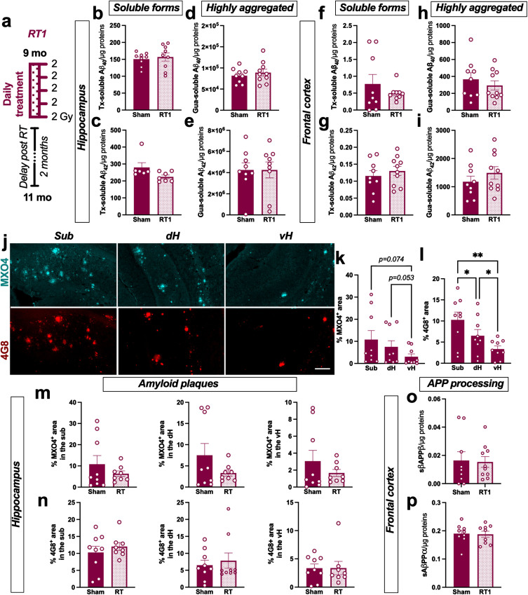
Results: A daily treatment did not affect amyloid and reduced only microglial-mediated inflammation markers, the opposite of the results obtained in our previous male study. Moreover, altered fractionations (2 Gy×5 weekly fractions or 10 fractions of 1 Gy delivered twice a week) did not influence the amyloid load or neuroinflammatory response in females.

Conclusions: A daily treatment consequently appears to be the most efficient for AD. This study also shows that the anti-amyloid and anti-inflammatory response to LD-RT are, at least partly, two distinct mechanisms. It also emphasizes the necessity to assess the sex impact when evaluating responses in ongoing pilot clinical trials testing LD-RT against AD.

Keywords: Alzheimer’s disease; amyloid; low-dose radiation therapy; microglial response.


Conflict of interest statement

The authors have no conflict of interest to report.

Figures

Fig. 1
Fig. 1
LD-RT does not reduce amyloid load in early AD females. a) Female TgF344-AD were bilaterally treated by X-ray radiation (2 Gy×5 fractions delivered daily; RT1) at 9-month-old, modeling an early AD stage, and analyzed 2 months later. Sham-treated TgAD rats and WT were only anesthetized. Concentration of Aβ40 (b) and Aβ42 (c) measured in the triton (Tx) soluble fraction of the hippocampus by ELISA. Concentration of Aβ40 (d) and Aβ42 (e) measured in Gua-soluble fraction of the hippocampus by ELISA. (f–i) Concentration of the different Aβ peptides in the frontal cortex by ELISA. (j) Representative images of amyloid plaques labelled with Methoxy-XO4 (MXO4; blue) or 4G8 antibody (red) in hippocampal subregions in a sham treated TgAD rat. Scale bar = 150 μm. Distribution of MXO4+ (k) and 4G8+ (l) plaques in the hippocampal subregions (Sub, subiculum; dH, dorsal hippocampus; vH, ventral hippocampus) in sham animals. Quantification of MXO4+ (m) and 4G8+ (n) plaques in the different hippocampal subregions in sham and RT1-treated rats. Concentration of sAβPPβ (o) and sAβPPα (p) in the Tx-soluble fraction of the frontal cortex measured by ELISA. Unpaired t-test or One-way ANOVA and Tukey’s multiple comparisons test. *p < 0.005, **p < 0.01.
Fig. 2
Fig. 2
LD-RT reduces microglial inflammation markers in early AD females. Quantification of astrocytic markers including GFAP (a), STAT3α (b), secreted CLUSTERIN (sCLU) (c), and SERPINA3 N (d) levels by western blot in the frontal cortex. Quantification of GFAP (e), STAT3α (f), secreted CLUSTERIN (sCLU) (g), and SERPINA3N (h) levels by western blot in the hippocampus. Data are normalized to GAPDH levels. One-way ANOVA and Tukey’ multiple comparisons test. i) Representative images of GFAP (magenta) staining in the subiculum (sub), dorsal hippocampus (dH) and ventral hippocampus (vH) of WT and sham treated TgAD rats. Scale bar = 100 μm. j) Quantification of the GFAP density (% positively stained area) in the Sub, dH and vH in sham-treated TgAD rats. k) Quantification of the GFAP density (% positively stained area) in the different groups in the sub, dH and vH. l) Representative images of CD68 (grey; merged with DAPI in blue) staining in the sub, dH and vH of WT and sham treated TgAD rats. Scale bar = 100 μm. m) Quantification of the number of CD68+ cells/μm2 in sham-treated TgAD rats. n) Quantification of the number of CD68+ cells/μm2 in the different groups in the sub, dH and vH. One-way ANOVA and Tukey’s multiple comparisons test or Kruskal-Wallis and Dunn’s multiple comparisons test. (o–q) mRNA levels quantified by qPCR in the cortex of animals. Two-way ANOVA (group and gene as between factors) and Tukey’s multiple comparisons test. # or *p < 0.05, **p < 0.01, ***p < 0.001.
Fig. 3
Fig. 3
Altered fractionations of radiation do not improve anti-amyloid effects in females. a) Female TgF344-AD rats were bilaterally treated by X-ray radiation (2 Gy×5 fractions delivered once-a-week; RT2 or 1 Gy×10 fractions delivered twice a week; RT3) at 9-month-old and analyzed 2 months later. Concentration of Aβ40 (b) and Aβ42 (c) measured in the triton (Tx) soluble fraction of the hippocampus by ELISA. Concentration of Aβ40 (d) and Aβ42 (e) measured in Gua-soluble fraction of the hippocampus by ELISA. (f–i) Concentration of different Aβ peptides in the frontal cortex by ELISA. Amyloid plaque density in the hippocampus stained using Methoxy-XO4 (MXO4+; j) or 4G8 antibody (k). Concentration of sAβPPβ (l) and sAβPPα (m) in the Tx-soluble fraction of the frontal cortex measured by ELISA. One-way ANOVA or Kruskal-Wallis and Dunn’s comparisons test. *p < 0.05.
Fig. 4
Fig. 4
Altered fractionations of radiation do not reduce inflammation markers in females. (a) Representative western blot images in the frontal cortex. Quantification of GFAP (b), STAT3α (c), secreted CLUSTERIN (sCLU) (d), and SERPINA3N (e) levels by western blot in the frontal cortex. (f) Representative western blot images in the hippocampus. Quantification of GFAP (g), STAT3α (h), sCLU (i), and SERPINA3N (j) levels by western blot in the hippocampus. Data are normalized to GAPDH levels. One-way ANOVA and Tukey’s multiple comparisons test. (k) Quantification of the GFAP density (% positively stained area) in the subiculum (sub), dorsal hippocampus (dH) and ventral hippocampus (vH) in the different groups. (l) Quantification of the number of CD68+ cells/μm2 in the hippocampal subregions. One-way ANOVA and Tukey’s multiple comparison test or Kruskal-Wallis and Dunn’s multiple comparisons test. (m–o) mRNA levels quantified by qPCR in the cortex of animals. Two-way ANOVA (group and gene as between factors) and Tukey’s multiple comparison test. Correlation of GFAP density and % of MXO4+ plaques (p) or % of 4G8+ plaques (q) in the entire hippocampus. Correlation of the number of CD68+ cells/μm2 and the % of MXO4+ plaques (r) or the % of 4G8+ plaques (s) in the entire hippocampus. Different symbols represent the subregions (Sub = magenta; dH = blue; vH = black). The Pearson coefficient (r) and the p value are indicated for the entire hippocampus. *p < 0.05, **p < 0.01, ***p < 0.001.
Fig. 5
Fig. 5
LD-RT does not influence behavioral performances of TgAD rats. (a) The spatial working memory of rats was assessed 2 months after the last session of LD-RT using the alternative Y maze test. One-way ANOVA. (b) Quantification of the general locomotion of animals in the open field after LD-RT. Kruskal-Wallis test.

Similar articles

Cited by

References

    1. Dar NJ, Glazner GW (2020) Deciphering the neuroprotective and neurogenic potential of soluble amyloid precursor protein alpha (sAPPα). Cell Mol Life Sci 77, 2315–2330. - PMC - PubMed
    1. Ben Haim L, Carrillo-de Sauvage M-A, Ceyzériat K, Escartin C (2015) Elusive roles for reactive astrocytes in neurodegenerative diseases. Front Cell Neurosci 9, 278. - PMC - PubMed
    1. Leng F, Edison P (2021) Neuroinflammation and microglial activation in Alzheimer disease: Where do we go from here? Nat Rev Neurol 17, 157–172. - PubMed
    1. Monterey MD, Wei H, Wu X, Wu JQ (2021) The many faces of astrocytes in Alzheimer’s disease. Front Neurol 12, 619626. - PMC - PubMed
    1. Ferretti MT, Iulita MF, Cavedo E, Chiesa PA, Schumacher Dimech A, Santuccione Chadha A, Baracchi F, Girouard H, Misoch S, Giacobini E, Depypere H, Hampel H, Women’s Brain Project and the Alzheimer Precision Medicine Initiative (2018) Sex differences in Alzheimer disease – the gateway to precision medicine. Nat Rev Neurol 14, 457–469. - PubMed

Publication types