. 2024 Jun 11;8(11):2660-2674.
doi: 10.1182/bloodadvances.2023011778.

Semaphorin 7A coordinates neutrophil response during pulmonary inflammation and sepsis

Affiliations

Semaphorin 7A coordinates neutrophil response during pulmonary inflammation and sepsis

Tiago Granja et al. Blood Adv. .

Abstract

Pulmonary defense mechanisms are critical for host integrity during pneumonia and sepsis. This defense is fundamentally dependent on the activation of neutrophils during the innate immune response. Recent work has shown that semaphorin 7A (Sema7A) holds significant impact on platelet function, yet its role on neutrophil function within the lung is not well understood. This study aimed to identify the role of Sema7A during pulmonary inflammation and sepsis. In patients with acute respiratory distress syndrome (ARDS), we were able to show a correlation between Sema7A and oxygenation levels. During subsequent workup, we found that Sema7A binds to the neutrophil PlexinC1 receptor, increasing integrins, and L-selectin on neutrophils. Sema7A prompted neutrophil chemotaxis in vitro and the formation of platelet-neutrophil complexes in vivo. We also observed altered adhesion and transmigration of neutrophils in Sema7A-/-animals in the lung during pulmonary inflammation. This effect resulted in increased number of neutrophils in the interstitial space of Sema7A-/- animals but reduced numbers of neutrophils in the alveolar space during pulmonary sepsis. This finding was associated with significantly worse outcome of Sema7A-/- animals in a model of pulmonary sepsis. Sema7A has an immunomodulatory effect in the lung, affecting pulmonary sepsis and ARDS. This effect influences the response of neutrophils to external aggression and might influence patient outcome. This trial was registered at www.ClinicalTrials.gov as #NCT02692118.


Conflict of interest statement

Conflict-of-interest disclosure: The authors declare no competing financial interests.

Figures

None
Graphical abstract
Figure 1.
Figure 1.
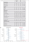
Clinical Sema7A values correlate with leukocyte count and clinical oxygenation values in patients with ARDS. Demographic data and samples of patients undergoing elective surgery with postoperative ventilation and ICU stays and patients admitted to the ICU for ARDS with severe pulmonary inflammation who were matched with propensity score. (A) Demographic data, ICU scores, and laboratory values for both patient groups are presented as means ± standard deviations, with values compared using the Wilcoxon rank-sum test. Significant values are set in bold. (B) The correlation of various laboratory values, ventilation parameters, and oxygenation values with serum levels of Sema7A is depicted. Pearson r and the lower and upper limits of the 95% confidence interval are shown. Significant correlations are highlighted in red. BE, Base excess; ICU, intensive care unit; INR, International Normalized Ratio; LDH, Lactate dehydrogenase.
Figure 2.
Figure 2.
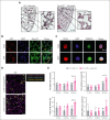
Sema7A is required for neutrophil adhesion and migration during inflammation. (A) Histological cross-sections and magnifications of lung tissue from WT and Sema7A−/− mice 24 hours after the instillation of 4 × 107 cells of K pneumoniae (scale bar, 200 μm). (B) IF staining of Sema7A (green) and vWF (red) in endothelial cells of murine lung tissue and nuclear staining with DAPI (4′,6-diamidino-2-phenylindole; blue; scale bar, 20 μm). (C) IF staining showing Sema7A expression (green) on the surface of human CD45-marked PMNs, (red) treated with NaCl or fMLP for 15 minutes (scale bar, 10 μm). (D) Representative video images of PNCs in murine lungs after LPS instillation with additional recombinant Sema7A or IgG2A-Fc (controls) treatment after 30 minutes (scale bar, 30 μm). (E) Total neutrophil area coverage, total and adhesive platelet area coverage, and the fractions of PNCs formed in the lung as determined via intravital confocal microscopic analysis of the lung in WT mice instilled with 5 μg/g BW LPS, with or without additional treatment with recombinant Sema7A (the data are the mean ± SD). ∗P < .05; ∗∗P < .01; ∗∗∗P < .001 as indicated. Fc, control fragment; IF, immunofluorescence; vWF, von Willebrand Factor.
Figure 3.
Figure 3.
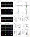
Sema7A binds to neutrophil PlexinC1 and influences neutrophil chemotaxis. Stained neutrophils isolated from saline (NaCl)- or LPS–treated WT and Sema7A−/− mice 4 hours after incubation. (A) Expression of Sema7A (red) and CD29 (green) on PMNs harvested from WT mice treated with NaCl or LPS (scale bar, 10 μm). (B) Expression of Sema7A (red) and EGFR (green) on PMNs harvested from WT mice treated with NaCl or LPS. No protein colocalization was visible in the merged pictures in either condition (scale bar, 10μm). (C) Expression of Sema7A (red) and PlexinC1 (green) on PMNs harvested from WT mice treated with NaCl or LPS. Sema7A expression is highly increased in LPS–treated mice, and the merged pictures show a strong interaction between Sema7A and PlexinC1 (scale bar, 10 μm). (D) Surface PMN expression of Sema7A (red) and PlexinC1 (green) in Sema7A−/− mice after the injection of exogenous recombinant Sema7A or IgG-Fc (control) after LPS or NaCl (controls) inhalation. Strong binding of exogenous Sema7A to Sema7A−/− PMNs was observed in LPS inhalation group. Multiple acquisitions of stained cells were analyzed from independently performed triplicate experiments (scale bar, 10 μm). (E) Human PMNs were subjected to different stimuli in bidirectional chemotactic chambers. Acquired time lapse videos over a 3-hour period were analyzed. Representative plots of PMN chemotactic tracks toward NaCl (control; red), fMLP (green), recombinant human SEMA7A (recSema7A; blue), or recSema7A together with antibodies against human PlexinC1 (anti-PLEXINC1; gray). (F-I) Comparison of the chemotaxis parameters forward migration index (FMI), Euclidean distance under the aspect of the direction, PMN velocity, and accumulated PMN distance. (J) PMN binding affinity was indicated by APClabeled fibrinogen on the surface of Ly6G-positive PMNs, as analyzed by FACS. The EDTA group was the internal negative control to measure the baseline autofluorescence, the untreated group was the fibrinogen-negative control, TNF-α was used as a potent PMN stimulator, and treatment of PMNs with recombinant SEMA7A before APClabeled fibrinogen represented the fibrinogen binding target group of interest. The fibrinogen-APC MFI was normalized and is displayed as a percentage. (K) PMN binding affinity was indicated by PerCP-labeled ICAM-1 on the surface of Ly6G-positive PMNs by FACS. The untreated group was the ICAM-1–negative control, TNF-α was used as a potent PMN stimulator, and treatment of PMNs with recombinant SEMA7A before PerCP-labeled ICAM-1 represented the ICAM-1 binding target group of interest. CD11b antibody treatment was used as a control for the inactivation of ICAM-1 binding. The ICAM-1 PerCP MFI was normalized and is displayed as a percentage. In (F-K), all group comparisons were performed by unpaired 2-tailed Student t tests (the data are the mean ± SD); ∗P < .05; ∗∗P < .01; ∗∗∗P < .001 as indicated. MFI, Mean Fluorescence Intensity.
Figure 4.
Figure 4.
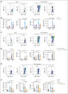
Essential neutrophil integrins are influenced by SEMA7A. Human PMNs were incubated with NaCl, 10 ng/mL TNF-α, or 2 μg/mL recSEMA7A for 15 minutes before proteomics analysis. The acquired raw data were analyzed after normalization. To analyze the samples, a 1-Factorial linear model was fitted with LIMMA, resulting in a 2-sided t test or F test based on moderated statistics. All presented P values were adjusted for multiple analyses by controlling the false discovery rate according to Benjamini and Hochberg. Proteins were defined as differential when |logFC| >.5 and an adjusted P value <.05 from triplicate experiments. (A) Expression of neutrophil surface lectin proteins and membrane integrin proteins from harvested samples. (B) Expression of intracellular neutrophil Rho/Ras GTPases, mitogen-associated kinases, adapter proteins, and detoxifying enzymes. (C) PMN surface expression of CD11b after 15 minutes of incubation with recSEMA7A or TNF-α. Measurement was performed by FACS, and the MFI (PE) was normalized to the highest measured value. (D) PMN surface expression of PSGL-1 after 15 minutes of incubation with recSEMA7A or TNF-α. (E) PMN surface expression of CD11a after 15 minutes of incubation with recSEMA7A or TNF-α. (F) PMN surface expression of CD62L after 15 minutes of incubation with recSEMA7A or TNF-α. Measurement was performed by FACS, and the MFI (BV510) was normalized to the highest measured value. Multiple cells were analyzed from independently performed experiments in triplicate. Group comparisons were performed by unpaired 2-tailed Student t tests (the data are the mean ± SD). ∗P < .05; ∗∗P < .01; ∗∗∗P < .001 as indicated. MFI, Mean Fluorescence Intensity; PE, Phycoerythrin.
Figure 5.
Figure 5.
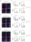
Tissue-specific expression of Sema7A controls neutrophil migration in response to inflammation. Intravital microscopic analysis of murine cremaster tissue after IV. LPS stimulation shows the role of Sema7A expression in different cells during inflammation. (A) Representative video images of the microvasculature of LysMCre+Sema7AloxP/loxP mice and littermate controls exposed to LPS for 15 minutes compared with the baseline control (0 min; scale bar, 50 μm). (B) Cell speed, transmigration, transmigration distance, and stationary PMNs in LysMCre+Sema7AloxP/loxP and littermate controls were analyzed by intravital microscopy after exposure to LPS for 15 minutes and compared with the baseline control (0 min). (C) Representative video images of the microvasculature of PF4Cre+Sema7AloxP/loxP mice and littermate controls exposed to LPS for 15 minutes compared with the baseline control (0 min; scale bar, 50 μm). (D) Cell speed, transmigration, transmigration distance, and stationary PMNs of PF4Cre+Sema7AloxP/loxP mice and littermate controls were analyzed by intravital microscopy after exposure to LPS for 15 minutes and compared with the baseline control (0 min; scale bar, 50 μm). (E) Representative video images of the microvasculature of Tie2Cre+Sema7AloxP/loxP mice and littermate controls exposed to LPS for 15 minutes and compared with the baseline control (0 min). (F) Cell speed, transmigration, transmigration distance, and stationary PMNs in Tie2Cre+Sema7AloxP/loxP and littermate controls were analyzed by intravital microscopy after exposure to LPS for 15 minutes and compared with the baseline control (0 min). Triplicate experiments were performed, and multiple cells were tracked for 15 to 20 minutes after LPS incubation over periods of 10 seconds at 90 fps. From the acquired videos, cells were tracked manually, and relevant group comparisons were performed by unpaired 2-tailed Student t tests (the data are the mean ± SD). ∗P < .05; ∗∗P < .01; ∗∗∗P < .001 as indicated, (arrows mark PNCs).
Figure 6.
Figure 6.
Activation of neutrophils and PNC formation is Sema7A dependent. Murine blood was collected from WT and Sema7A−/− mice after LPS inhalation and analyzed by flow cytometry. (A) Representative color dot blots of PNCs (Ly6G+/CD42b+ events) in WT and Sema7A−/− blood from NaCl (control) or LPS–inhaled mice. (B) PNC formation, platelet effector glycoprotein 2b/3a (GP2b/3a) expression (antibody clone JON/A MFI), PMN activity marker CD11b (MFI) expression, and platelet activity marker CD42b (MFI) expression were assessed by flow cytometry in the mice described in panel A. (C) Representative dot blots of PNCs (Ly6G+/CD42b+ events) in the blood of WT and Sema7A−/− mice treated with recombinant Sema7A (recSema7A) after NaCl (control) or LPS inhalation. (D) PNC formation, platelet effector GPIIb/IIIa expression (antibody clone JON/A MFI), PMN activity marker CD11b (MFI) expression, and platelet activity marker CD42b (MFI) expression were assessed by flow cytometry in the mice described in panel C. (E) Representative dot blots of PNCs (Ly6G+/CD42b+ events) in the blood of WT mice that were untreated or treated with IgG or the Sema7A-blocking antibody (anti-Sema7A) after LPS inhalation compared with mice without conditioning (Sham). (F) PNC formation, platelet effector GPIIb/IIIa expression (antibody clone JON/A MFI), PMN activity marker CD11b (MFI) expression, and platelet activity marker CD42b (MFI) expression were assessed by flow cytometry in the mice described in panel E. Group comparisons were performed by unpaired 2-tailed Student t tests (the data are the mean ± SD). ∗P < .05; ∗∗P < .01; ∗∗∗P < .001 as indicated. IgG, immunoglobulin G; MFI, Mean Fluorescence Intensity.
Figure 7.
Figure 7.
Sema7A is crucial for pulmonary defense against Klebsiella-induced pneumonia. In a murine model of bacterial-induced lung injury, 4 × 107 gram-negative K pneumoniae was administered by intratracheal instillation directly into the lungs of WT and Sema7A−/− mice. Measurements of PMN counts on the endothelial surface (A), in the interstitial space (B), in the BAL (C), and PNC numbers (D) per tissue section (magnification, 1000×) 24 hours after Klebsiella instillation in histological lung sections of WT and Sema7A−/− mice (n = 3 per group on 3 different layers). The proinflammatory cytokines TNF-α (E), IL-6 (F), IL-1β (G), and myeloperoxidase activity (H) within the BAL of WT and Sema7A−/− mice. (I) Histological sections demonstrating the quantity of Klebsiella, the alveolar inflammation (H&E staining), and PNC debris (PNC-specific staining) 24 hours after instillation (scale bar, 50 μm; magnification, 1000×). Colony forming units in BALF (J) and blood (K) taken 24 hours after Klebsiella instillation and incubated on nutrient agar plates for 24 h. (L) Representative images of cultured bacteria and (M) counts per tissue sections of K pneumoniae. (N) Representative images of H&E stained sections focusing on lung tissue injury 24 hours after Klebsiella instillation. (scale bar, 50 μm; magnification, 1000×). (O) Thickness of alveolar wall in tissue sections of WT and Sema7A−/− mice 24 hours after K pneumoniae instillation (n = 3 per group; 10 random fields of view per mouse). (P) Survival curves of WT and Sema7A−/− animals after the instillation of 4 × 107 cells of K pneumoniae (n ≥ 6 per group). Group comparisons were performed by unpaired 2-tailed Student t tests; the data are the mean ± SD. For statistical comparisons of survival, the Gehan-Breslow-Wilcoxon test and the log-rank (Mantel-Cox) test were performed. ∗P < .05; ∗∗P < .01; ∗∗∗P < .001 as indicated. (Q) Schematic drawing of the role of Sema7a in pulmonary infection and defense. In pulmonary hemostasis, Sema7A is expressed on neutrophils and other tissues (left). During pulmonary infection Sema7A gains pathophysiological importance. Sema7A binds to Plexin C1, activates neutrophils, and increases the expression of integrins and L-selectin on their surface. This is important for a coordinated immunological response and the defense of the lung. BALF, bronchoalveolar fluid; H&E, hematoxylin-eosin; IL-6, interleukin-6.

References

    1. Singer M, Deutschman CS, Seymour CW, et al. The third international consensus definitions for sepsis and septic shock (Sepsis-3) JAMA. 2016;315(8):801–810. - PMC - PubMed
    1. Prina E, Ranzani OT, Torres A. Community-acquired pneumonia. Lancet. 2015;386(9998):1097–1108. - PMC - PubMed
    1. Holt PG, Strickland DH, Wikstrom ME, Jahnsen FL. Regulation of immunological homeostasis in the respiratory tract. Nat Rev Immunol. 2008;8(2):142–152. - PubMed
    1. Potey PM, Rossi AG, Lucas CD, Dorward DA. Neutrophils in the initiation and resolution of acute pulmonary inflammation: understanding biological function and therapeutic potential. J Pathol. 2019;247(5):672–685. - PMC - PubMed
    1. Yoshida K, Kondo R, Wang Q, Doerschuk CM. Neutrophil cytoskeletal rearrangements during capillary sequestration in bacterial pneumonia in rats. Am J Respir Crit Care Med. 2006;174(6):689–698. - PMC - PubMed

Publication types

Associated data