. 2024 Mar 15;10(11):eadk2542.
doi: 10.1126/sciadv.adk2542. Epub 2024 Mar 15.

Mono-UFMylation promotes misfolding-associated secretion of α-synuclein

Affiliations

Mono-UFMylation promotes misfolding-associated secretion of α-synuclein

Lihui Wang et al. Sci Adv. .

Abstract

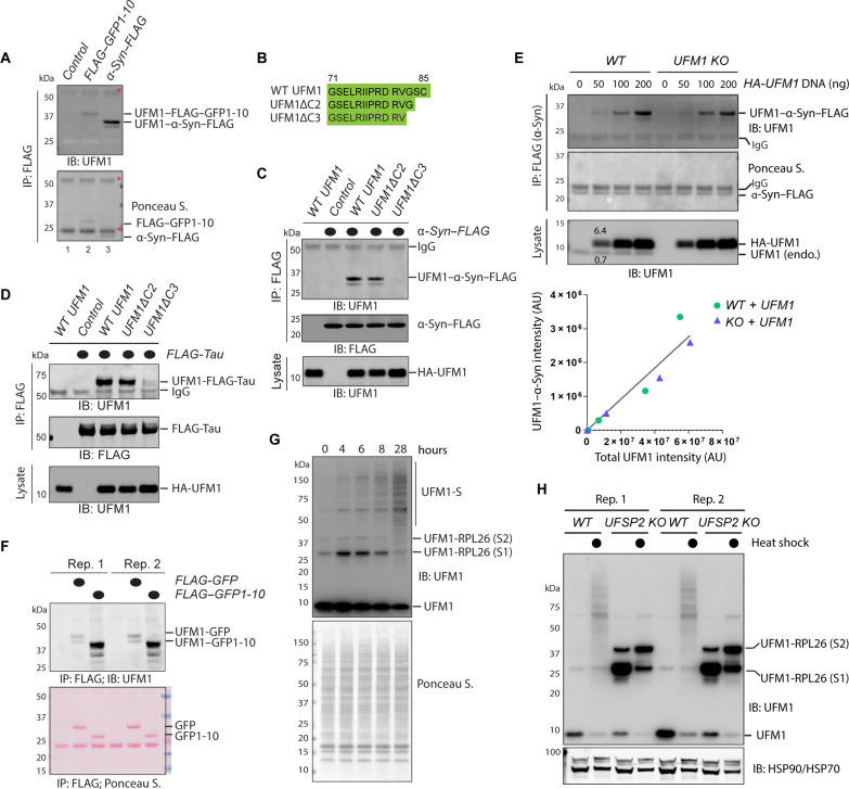
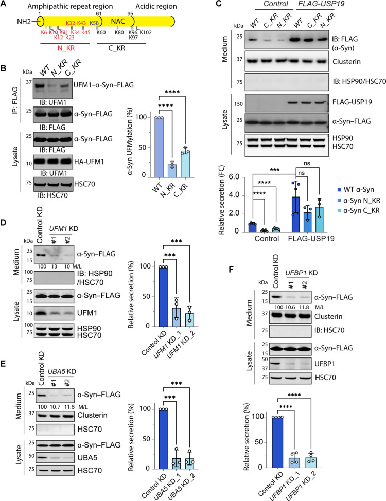
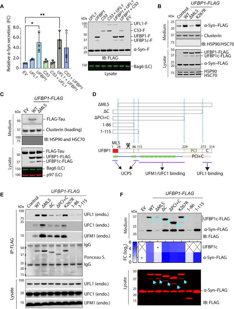
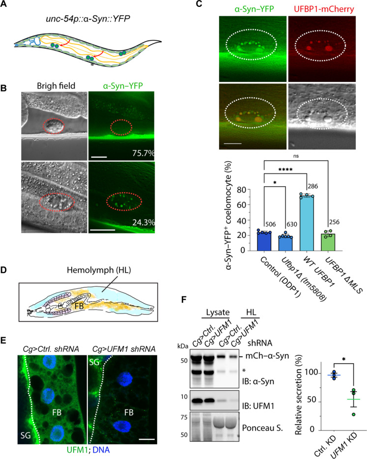
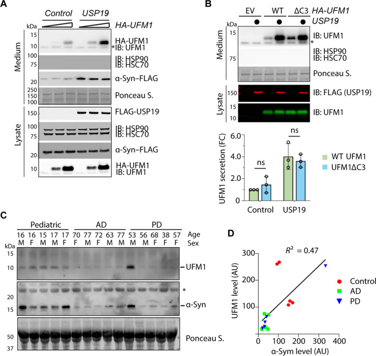
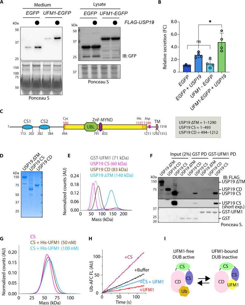
Stressed cells secret misfolded proteins lacking signaling sequence via an unconventional protein secretion (UcPS) pathway, but how misfolded proteins are targeted selectively in UcPS is unclear. Here, we report that misfolded UcPS clients are subject to modification by a ubiquitin-like protein named ubiquitin-fold modifier 1 (UFM1). Using α-synuclein (α-Syn) as a UcPS model, we show that mutating the UFMylation sites in α-Syn or genetic inhibition of the UFMylation system mitigates α-Syn secretion, whereas overexpression of UFBP1, a component of the endoplasmic reticulum-associated UFMylation ligase complex, augments α-Syn secretion in mammalian cells and in model organisms. UFM1 itself is cosecreted with α-Syn, and the serum UFM1 level correlates with that of α-Syn. Because UFM1 can be directly recognized by ubiquitin specific peptidase 19 (USP19), a previously established UcPS stimulator known to associate with several chaperoning activities, UFMylation might facilitate substrate engagement by USP19, allowing stringent and regulated selection of misfolded proteins for secretion and proteotoxic stress alleviation.


Figures

Fig. 1.
Fig. 1.. Misfolded MAPS substrates are mono-UFMylated.
(A) UFMylation of GFP1-10 and α-Syn in HEK293T cells. Cells transfected with HA-tagged UFM1 together with either an empty vector control or FLAG–GFP1-10 or α-Syn–FLAG were subject to immunoprecipitation (IP) by FLAG beads under denaturing conditions. Precipitated proteins were analyzed by immunoblotting (IB). Asterisks indicate immunoglobulin G (IgG). (B) The amino acid sequence of the C terminus of WT UFM1 and the mutants used in the study. (C) UFMylation of α-Syn requires G83 of UFM1. As in (A), except that cells were transfected with the indicated plasmids. Where indicated, 15% of the total lysates were analyzed together with the immunoprecipitated samples. (D) UFMylation of Tau in HEK293T cells. As in (C), except that FLAG-tagged Tau was used. (E) The level of UFMylated α-Syn correlates with UFM1 concentrations in cells. α-Syn–FLAG immunoprecipitated from HEK293T cells transfected with the indicated plasmids were analyzed by immunoblotting. The graph shows a linear correlation between the level of UFMylated α-Syn and UFM1. AU, arbitrary units. (F) UFMylation of GFP1-10 is more efficient than full-length GFP. Cells transfected with HA-UFM1 together with the indicated GFP variants were analyzed as in (A). Shown is a blot with two biological repeats (Rep.). (G) Heat shock induces protein UFMylation. HEK293T cells treated at 43°C for the indicated time points were lysed in sample buffer and analyzed by immunoblotting with UFM1 antibodies. UFM1-S, heat-induced UFMylated substrates. (H) Protein UFMylation under the heat shock condition is not regulated by UFSP2. Whole-cell extracts from untreated or heat-treated (16 hours, indicated by filled circles) WT or UFSP2 CRISPR KO cells were analyzed by immunoblotting. Note that UFSP2 KO increases ribosome UFMylation but inhibits heat-induced UFMylation.
Fig. 2.
Fig. 2.. UFMylation of α-Syn promotes its secretion.
(A) A schematic diagram showing the lysine positions in α-Syn and the mutants tested. (B) Mono-UFMylation of α-Syn can occur on multiple lysine residues. HEK293T cells were transfected with HA-UFM1 together with the indicated α-Syn lysine mutants and then subject to immunoprecipitation and immunoblotting. The graph shows the quantification of three biological repeats. Error bars indicate means ± SD. ****P < 0.0001 by one-way analysis of variance (ANOVA) with Dunnett’s multiple comparison test. (C) Reduced secretion of α-Syn lysine mutants. Immunoblotting analysis of condition medium and lysates from HEK293T cells transfected with the indicated plasmids. The graph shows the quantification of the relative ratio of α-Syn in medium normalized to that in the corresponding lysate (M/L). The dots indicate the number of biological repeats. FC, fold change. Error bars indicate means ± SD. ****P < 0.0001; ***P < 0.001; ns, not significant by one-way ANOVA. (D) Knockdown (KD) of UFM1 reduces α-Syn secretion. Conditioned medium and lysates from cells transfected with α-Syn–FLAG together with the indicated siRNAs were analyzed by immunoblotting. The numbers and the graph show the relative ratio of medium α-Syn versus that in lysates (M/L). Error bars indicate means ± SD. ***P < 0.001 by one-way ANOVA, n = 3 biological repeats. (E) Knockdown of UBA5 reduces α-Syn secretion. Same as (D), except that two UBA5-specific siRNAs were used. Error bars indicate means ± SD. ***P < 0.001 by one-way ANOVA, n = 3 biological repeats. (F) Knockdown of UFBP1 reduces α-Syn secretion. Same as (D), except that two UFBP1-targeting siRNAs were used and that n = 4 biological repeats. Error bars indicate means ± SD. ****P < 0.001 by one-way ANOVA.
Fig. 3.
Fig. 3.. Overexpression of UFBP1 induces α-Syn secretion in mammalian cells.
(A) Overexpression of UFBP1 stimulates α-Syn secretion. Conditioned medium and lysates from HEK293T cells transfected with α-Syn–FLAG together with the indicated plasmids were analyzed by immunoblotting. F, FLAG tag; C53, CDK53RAP1; EV, empty vector control. The graph shows secreted α-Syn normalized by α-Syn in cell lysates (right). Error bars indicate means ± SD. *P < 0.05; **P < 0.01 by unpaired two-tailed t test. n = 3 biological repeats. (B) As in (A), except that α-Syn–FLAG was cotransfected with the indicated plasmids. (C) As in (A), except that FLAG-tagged Tau was expressed together with the indicated UFBP1 variants. LC, loading control. (D) A schematic diagram shows the UFBP1 variants tested in (E) and (F). The bottom graph shows the predicted secondary and domain structure of UFBP1. Domains involved in known protein-protein interactions are marked. (E) The interactions of UFBP1 variants with the endogenous UFM1, UFC1, and UFL1 were analyzed by coimmunoprecipitation, followed by immunoblotting. (F) UFBP1 is cosecreted with α-Syn. Shown is a representative immunoblotting analysis of conditioned medium and lysates from cells transfected with α-Syn–FLAG together with the indicated UFBP1 variants. Note that a cleaved UFBP1 fragment (UFBP1c-FLAG) was cosecreted with α-Syn with the exception for UFBP1 1-86 and 1-115. The heatmap shows the relative secretion of UFBP1c and α-Syn. Asterisks indicate the samples used for fold change normalization. n = 2 biological repeats. X = not detected. Arrows denote UFBP1c.
Fig. 4.
Fig. 4.. The UFMylation system facilitates α-Syn secretion in model organisms.
(A) A schematic diagram of the worm-based secretion assay. (B) α-Syn is secreted from body wall muscle cells and internalized by coelomocytes in C. elegans. Worms bearing a Venus (YFP)–tagged α-Syn were imaged by a fluorescence microscope. The images represent WT worms with two different phenotypes. Scale bars, 10 μm. (C) UFBP1 promotes α-Syn secretion in C. elegans. Top: Representative images of coelomocyte-accumulated α-Syn–YFP and UFBP1-mCherry. The graph shows the quantification of worms of the indicated genotypes with α-Syn–YFP–positive (+) coelomocyte. Dots indicate the number of repeats, whereas the numbers indicate total worms counted. Error bars indicate means ± SD. *P < 0.05; ****P < 0.0001, by one-way ANOVA. Scale bar, 5 μm. (D) A schematic illustration of the fly UcPS model. FB, fat body; HL, hemolymph. (E) Immunostaining confirms fat body–specific knockdown of UFM1. Fat bodies isolated from third instar larvae of the indicated genotypes were stained with a UFM1 antibody (green) and 4′,6-diamidino-2-phenylindole (blue). Ctrl. shRNA, control shRNA. Note that the UFM1 signal is only specifically reduced in fat body. SG, salivary gland. Scale bar, 20 μm. (F) Fat body–specific knockdown of UFM1 reduces α-Syn secretion. Hemolymph and hemolymph-depleted larva lysates were analyzed by immunoblotting. The asterisk indicates a cleaved α-Syn species. The graph shows the quantification of three biological repeats. Error bars indicate means ± SD. *P < 0.05 by unpaired two-tailed t test. n = 3 biological repeats.
Fig. 5.
Fig. 5.. Endogenous UFM1 is cosecreted with α-Syn.
(A) USP19 promotes UFM1 secretion. Conditioned medium and lysates from HEK293T cells transfected with different amounts of HA-UFM1 together with an empty vector control or FLAG-USP19 were analyzed by immunoblotting. (B) The secretion of UFM1 does not require G83. The indicated UFM1 variants or an empty vector control were transfected together with (indicated by filled circles) or without USP19. The graph shows the quantification of three biological repeats. Error bars indicate means ± SD. (C) Endogenous UFM1 and α-Syn are detected in human serum. Serum from patients with Alzheimer’s disease (AD) and Parkinson’s disease (PD) were analyzed together with a group of pediatric serum. (D) Quantification of the UFM1 and α-Syn levels in human serum samples.
Fig. 6.
Fig. 6.. UFM1 binds and regulates USP19.
(A) USP19 promotes the secretion of UFM1-EGFP. Conditioned medium and lysates from cells transfected as indicated were analyzed by immunoblotting. (B) Quantification of the experiments shown in (A). Error bars indicate means ± SD. *P < 0.05 by unpaired two-tailed t test. n = 4 biological repeats. (C) A diagram showing the USP19 domain structure and the truncation variants tested. (D) A Coomassie blue–stained gel showing the purified proteins. (E) Mass photometry confirms the molecular mass of the purified proteins. (F) A GST pull-down (PD) assay confirms the interaction of USP19 CS with UFM1. (G) Mass photometry demonstrates the interaction of His-UFM1 with USP19 CS (50 nM). (H) USP19 CS stimulates the deubiquitinating activity of USP19 CD, which is antagonized by UFM1. The deubiquitinating activity of USP19 CD (20 nM) was measured together with either a buffer control, or USP19 CS (200 nM), or UFM1 (5 μM) or the combination of USP19 CS (200 nM) and UFM1 (5 μM). (I) A toggle switch model showing USP19 in two functional states, a UFM1-free DUB active, and a UFM1-bound DUB-inactive form.

Similar articles

Cited by

References

    1. Benham A. M., Protein secretion and the endoplasmic reticulum. Cold Spring Harb. Perspect. Biol. 4, a012872 (2012). - PMC - PubMed
    1. Rabouille C., Pathways of unconventional protein secretion. Trends Cell Biol. 27, 230–240 (2017). - PubMed
    1. Nickel W., Rabouille C., Unconventional protein secretion: Diversity and consensus. Semin. Cell Dev. Biol. 83, 1–2 (2018). - PubMed
    1. Steringer J. P., Lange S., Čujova S., Šachl R., Poojari C., Lolicato F., Beutel O., Müller H. M., Unger S., Coskun Ü., Honigmann A., Vattulainen I., Hof M., Freund C., Nickel W., Key steps in unconventional secretion of fibroblast growth factor 2 reconstituted with purified components. eLife 6, e28985 (2017). - PMC - PubMed
    1. New J., Thomas S. M., Autophagy-dependent secretion: Mechanism, factors secreted, and disease implications. Autophagy 15, 1682–1693 (2019). - PMC - PubMed

Publication types