. 2024 Mar 15;10(11):eadh4435.
doi: 10.1126/sciadv.adh4435. Epub 2024 Mar 15.

Increased genomic instability and reshaping of tissue microenvironment underlie oncogenic properties of Arid1a mutations

Affiliations

Increased genomic instability and reshaping of tissue microenvironment underlie oncogenic properties of Arid1a mutations

Alessandro D'Ambrosio et al. Sci Adv. .

Abstract

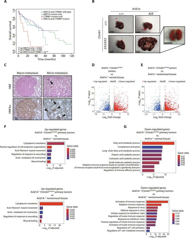
Oncogenic mutations accumulating in many chromatin-associated proteins have been identified in different tumor types. With a mutation rate from 10 to 57%, ARID1A has been widely considered a tumor suppressor gene. However, whether this role is mainly due to its transcriptional-related activities or its ability to preserve genome integrity is still a matter of intense debate. Here, we show that ARID1A is largely dispensable for preserving enhancer-dependent transcriptional regulation, being ARID1B sufficient and required to compensate for ARID1A loss. We provide in vivo evidence that ARID1A is mainly required to preserve genomic integrity in adult tissues. ARID1A loss primarily results in DNA damage accumulation, interferon type I response activation, and chronic inflammation leading to tumor formation. Our data suggest that in healthy tissues, the increased genomic instability that follows ARID1A mutations and the selective pressure imposed by the microenvironment might result in the emergence of aggressive, possibly immune-resistant, tumors.


Figures

Fig. 1.
Fig. 1.. ARID1A loss sensitizes to HCC formation.
(A) Experimental scheme. (B) Gross examination and histological and immunohistochemical analysis of wild-type and Arid1a−/− liver 5 months from NR1L3 agonist injection showing liver tumor formation (arrowheads). (C) Number of animals that developed tumors upon sporadic activation of NR1L3. (D) Volcano plot showing transcriptional alterations observed by RNA-seq analysis in Arid1a−/− compared to wild-type animals 30 days from NR1L3 agonist administration. (E) Bar plot showing overrepresented biological process (GO terms) of up-regulated genes in Arid1a−/− compared to wild-type animals 30 days from NR1L3 agonist administration. (F) Gross examination of wild-type and Arid1a−/− liver 15 months from tamoxifen injection showing liver tumor formation (arrowheads). N, nontumoral; T, tumor; H&E, hematoxylin and eosin.
Fig. 2.
Fig. 2.. ARID1A is largely dispensable for preserving enhancer-dependent transcriptional regulation.
(A) Heatmap of H3K27Ac and H3K4Me1 levels on ARID1A-bound distal regions in wild-type, Arid1a−/− and Arid1a/b−/− hepatocytes. The four sets of regions (quantiles) were generated on the basis of the H3K27Ac intensity ratio (Arid1a−/− versus wild type; n = 2, see Materials and Methods). (B) Heatmap and (C) density plot showing C/EBPα occupancy on ARID1A-bound distal regions in wild-type, Arid1a−/− hepatocytes. (D) Volcano plots showing transcriptional alterations observed by RNA-seq analysis in Arid1a−/− compared to wild type at 9 and 30 days from tamoxifen injection. (E) Volcano plot showing transcriptional alterations observed by RNA-seq analysis in Arid1a/b−/− liver compared to wild type at 9 days from tamoxifen injection. (F) Histological and immunohistochemical analysis of wild-type and Arid1a/b−/− liver samples. (G) UpSet plots representing the number of differentially expressed genes (Arid1a−/− versus wild type) regulated by the ARID1A-bound enhancers in the four different quantiles. Only intersections ≥ 3 are shown. Boxplots represent transcriptional changes of each specific set of genes in Arid1a−/− liver compared to wild type.
Fig. 3.
Fig. 3.. ARID1A loss induces interferon response activation.
(A) GO terms enriched between up-regulated genes in Arid1a−/− livers 9 days and 30 days from tamoxifen injection. (B) qPCR analysis of interferon target genes in purified hepatocytes 9 days from tamoxifen injection. (C) Common GO terms enriched in up-regulated genes in Arid1a−/− liver (this work), HCT116 (4), intestinal crypts (15), MCF7 (5), and liver from DDC-treated mice (28). (D) Immunohistochemistry analysis of wild-type and Arid1a−/− liver sections using anti ISG15, anti-CD8, and anti-IBA1 and quantification of CD8+ T cells and IBA1+ macrophages (unpaired t test, two-tailed).
Fig. 4.
Fig. 4.. ARID1A loss induces chronic inflammatory disease and DNA damage.
(A) Gross examination of wild-type and Arid1a−/− livers, 5 months from tamoxifen injection. (B) Histological and (C) immunohistochemical analysis of wild-type and Arid1a−/− liver 5 months from tamoxifen injection.
Fig. 5.
Fig. 5.. ARID1A loss promotes DNA damage accumulation.
(A) Immunohistochemical analysis of wild-type and Arid1a−/− liver 9 days from tamoxifen injection using CDKN1A, γH2AX, and KI67 antibodies. Quantifications are shown for three antibodies (unpaired t test, two-tailed). (B) Comet assay performed using wild-type and Arid1a−/− hepatocytes. The quantification of the tail moment is shown (n > 50 per condition. Unpaired t test, two-tailed). AU, arbitrary units. (C) Immunohistochemical analysis of gastric sections from Prom1CreERT2 wild-type and Arid1a−/− mice, 7 days after tamoxifen injection using CDKN1A and γH2AX antibodies. Quantification is shown for the two antibodies (unpaired t test, two-tailed). (D) Immunohistochemical staining of small intestine sections from Arid1Afl/fl (15).
Fig. 6.
Fig. 6.. ARID1A loss promotes micronuclei formation and sensitizes to ATR inhibitors.
(A) Immunohistochemical staining of wild-type and Arid1a−/− liver 9 days from tamoxifen injection using H3K27Me3 and γH2AX antibodies (arrowheads show micronuclei-containing cells) and quantification of micronuclei-containing cells. (B) Representative images of Arid1a-proficient and Arid1a-deficient hepatocytes during anaphase after 30 days from a single NR1L3 agonist injection. (C) 3D reconstruction obtained by 185 serial sections of nuclei (green) and micronuclei (purple) of hepatocyte in 16,650 μm3 of Arid1a−/− liver 9 days from tamoxifen injection. N, nucleus; MN, micronucleus. Two representative images in which the micronuclear envelope lesion (arrowheads) of the micronuclei present in the 3D reconstruction are visible. Scale bars, 1 μm. (D) Gene set enrichment analysis of differentially expressed genes in Fig. 1D performed using the CIN70 signature. (E) Representative images and cell viability of ARID1A wild-type and mutant human gastric cancer organoids treated with 10 μM berzosertib or ceralasertib for 48 hours. DMSO, dimethyl sulfoxide; NES, normalized enrichment score.
Fig. 7.
Fig. 7.. ARID1A-defective murine tumors recapitulate aggressive human HCC.
(A) Copy number alterations in an Arid1a−/− defective tumor. (B) SNV classification in Arid1a−/− murine tumors and human HCC from TCGA datasets. (C) Cosine similarity between human HCCs and Arid1a−/− murine tumors. (D) Mutational signature of Arid1a−/− tumors and relative contribution of known single base substitution (SBS) signatures (from COSMIC). (E) Table of representative genomic alterations of Arid1a−/− murine tumors. (F) β-Catenin staining of normal tissue and Ctnnb1 wild-type and Arid1a−/− tumors. mad, median absolute deviation.
Fig. 8.
Fig. 8.. Concomitant Arid1a and Ctnnb1 mutations promote metastatic liver tumors.
(A) Kaplan-Meier curves showing overall survival of patients with HCC harboring ARID1A and CTNNB1 mutations compared with single-mutant tumors. (B) Gross appearance of spontaneously develop tumors and lung metastasis in AlbCre-ERT2Arid1afl/flCtnnb1EX3/EX3 mice at 10 months of age. (C) Histological and immunohistochemical analysis of metastatic lesions in AlbCre-ERT2Arid1afl/flCtnnb1EX3/EX3 mice. (D) Volcano plot showing transcriptional alterations observed by RNA-seq analysis in AlbCre-ERT2Arid1afl/flCtnnb1EX3/EX3 primary tumors compared to wild-type normal tissues. (E) Volcano plot showing transcriptional alterations observed by RNA-seq analysis in AlbCre-ERT2Arid1afl/flCtnnb1EX3/EX3 primary tumor compared to Arid1a−/− nontumoral tissues. (F) Bar plot showing overrepresented biological process (GO terms) between up-regulated and (G) down-regulated genes in the AlbCre-ERT2Arid1afl/flCtnnb1EX3/EX3 primary tumors compared to wild-type normal tissues or Arid1a−/− nontumoral tissues.

References

    1. Centore R. C., Sandoval G. J., Soares L. M. M., Kadoch C., Chan H. M., Mammalian SWI/SNF chromatin remodeling complexes: Emerging mechanisms and therapeutic strategies. Trends Genet. 36, 936–950 (2020). - PubMed
    1. Cenik B. K., Shilatifard A., COMPASS and SWI/SNF complexes in development and disease. Nat. Rev. Genet. 22, 38–58 (2021). - PubMed
    1. Mittal P., Roberts C. W. M., The SWI/SNF complex in cancer - biology, biomarkers and therapy. Nat. Rev. Clin. Oncol. 17, 435–448 (2020). - PMC - PubMed
    1. Mathur R., Alver B. H., San Roman A. K., Wilson B. G., Wang X., Agoston A. T., Park P. J., Shivdasani R. A., Roberts C. W. M., ARID1A loss impairs enhancer-mediated gene regulation and drives colon cancer in mice. Nat. Genet. 49, 296–302 (2017). - PMC - PubMed
    1. Xu G., Chhangawala S., Cocco E., Razavi P., Cai Y., Otto J. E., Ferrando L., Selenica P., Ladewig E., Chan C., da Cruz Paula A., Witkin M., Cheng Y., Park J., Serna-Tamayo C., Zhao H. Y., Wu F., Sallaku M., Qu X., Zhao A., Collings C. K., D’Avino A. R., Jhaveri K., Koche R., Levine R. L., Reis-Filho J. S., Kadoch C., Scaltriti M., Leslie C. S., Baselga J., Toska E., ARID1A determines luminal identity and therapeutic response in estrogen-receptor-positive breast cancer. Nat. Genet. 52, 198–207 (2020). - PMC - PubMed

Publication types