

Model-based iterative reconstruction for direct imaging with point spread function encoded echo planar MRI
- PMID: 38490504
- PMCID: PMC11075760
- DOI: 10.1016/j.mri.2024.03.009
Model-based iterative reconstruction for direct imaging with point spread function encoded echo planar MRI
Abstract
Background: Echo planar imaging (EPI) is a fast measurement technique commonly used in magnetic resonance imaging (MRI), but is highly sensitive to measurement non-idealities in reconstruction. Point spread function (PSF)-encoded EPI is a multi-shot strategy which alleviates distortion, but acquisition of encodings suitable for direct distortion-free imaging prolongs scan time. In this work, a model-based iterative reconstruction (MBIR) framework is introduced for direct imaging with PSF-EPI to improve image quality and acceleration potential.
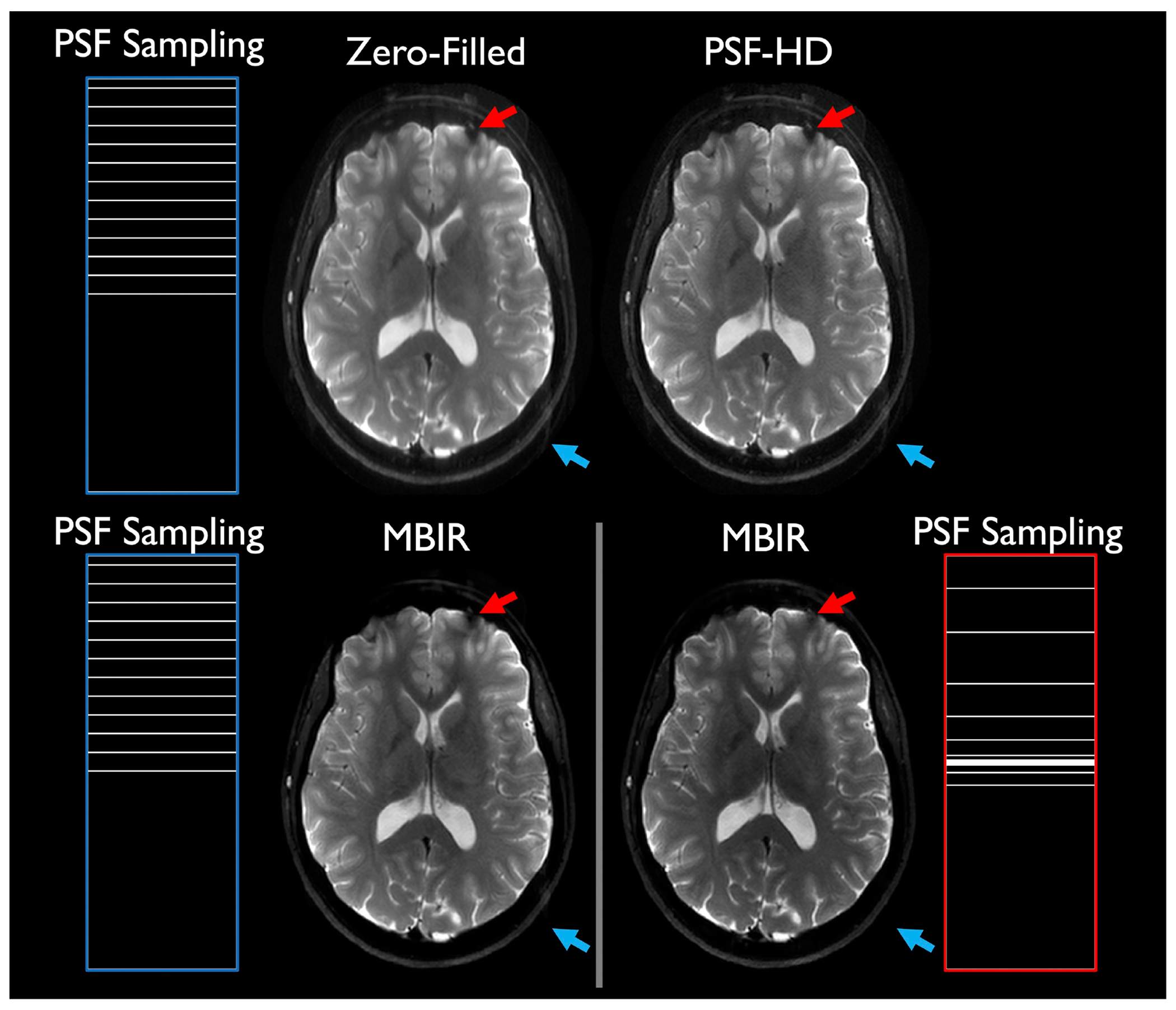
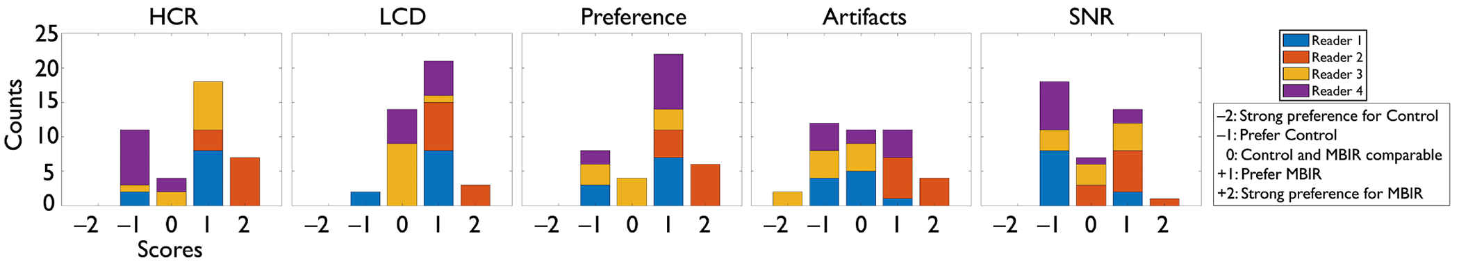
Methods: An MBIR platform was developed for accelerated PSF-EPI. The reconstruction utilizes a subspace representation, is regularized to promote local low-rankedness (LLR), and uses variable splitting for efficient iteration. Comparisons were made against standard reconstructions from prospectively accelerated PSF-EPI data and with retrospective subsampling. Exploring aggressive partial Fourier acceleration of the PSF-encoding dimension, additional comparisons were made against an extension of Homodyne to direct PSF-EPI in numerical experiments. A neuroradiologists' assessment was completed comparing images reconstructed with MBIR from retrospectively truncated data directly against images obtained with standard reconstructions from non-truncated datasets.
Results: Image quality results were consistently superior for MBIR relative to standard and Homodyne reconstructions. As the MBIR signal model and reconstruction allow for arbitrary sampling of the PSF space, random sampling of the PSF-encoding dimension was also demonstrated, with quantitative assessments indicating best performance achieved through nonuniform PSF sampling combined with partial Fourier. With retrospective subsampling, MBIR reconstructs high-quality images from sub-minute scan datasets. MBIR was shown to be superior in a neuroradiologists' assessment with respect to three of five performance criteria, with equivalence for the remaining two.
Conclusions: A novel image reconstruction framework is introduced for direct imaging with PSF-EPI, enabling arbitrary PSF space sampling and reconstruction of diagnostic-quality images from highly accelerated PSF-encoded EPI data.
Keywords: Echo planar imaging; Image reconstruction; Low-rank; Point spread function.
Copyright © 2024 Elsevier Inc. All rights reserved.
Conflict of interest statement
Declaration of competing interest MAB and JDT acknowledge the following interest: Mayo Clinic has licensed IP related to the compact 3T to GE Healthcare.
Figures


Similar articles
-
Tilted-CAIPI for highly accelerated distortion-free EPI with point spread function (PSF) encoding.Magn Reson Med. 2019 Jan;81(1):377-392. doi: 10.1002/mrm.27413. Epub 2018 Sep 5. Magn Reson Med. 2019. PMID: 30229562 Free PMC article.
-
Water/fat separation for distortion-free EPI with point spread function encoding.Magn Reson Med. 2019 Jul;82(1):251-262. doi: 10.1002/mrm.27717. Epub 2019 Mar 7. Magn Reson Med. 2019. PMID: 30847991
-
Rapid Geometry-Corrected Echo-Planar Diffusion Imaging at Ultrahigh Field: Fusing View Angle Tilting and Point-Spread Function Mapping.Magn Reson Med. 2022 Nov;88(5):2074-2087. doi: 10.1002/mrm.29360. Epub 2022 Jun 28. Magn Reson Med. 2022. PMID: 35762910
-
High-fidelity mesoscale in-vivo diffusion MRI through gSlider-BUDA and circular EPI with S-LORAKS reconstruction.Neuroimage. 2023 Jul 15;275:120168. doi: 10.1016/j.neuroimage.2023.120168. Epub 2023 May 13. Neuroimage. 2023. PMID: 37187364 Free PMC article.
-
Highly accelerated PSF-mapping for EPI distortion correction with improved fidelity.MAGMA. 2012 Jun;25(3):183-92. doi: 10.1007/s10334-011-0275-6. Epub 2011 Aug 4. MAGMA. 2012. PMID: 21814756
References
MeSH terms
Grants and funding
LinkOut - more resources
Full Text Sources
Research Materials
