. 2024 Mar 15;15(3):218.
doi: 10.1038/s41419-024-06594-w.

TGF-β-driven LIF expression influences neutrophil extracellular traps (NETs) and contributes to peritoneal metastasis in gastric cancer

Affiliations

TGF-β-driven LIF expression influences neutrophil extracellular traps (NETs) and contributes to peritoneal metastasis in gastric cancer

Fangbin Zhang et al. Cell Death Dis. .

Abstract

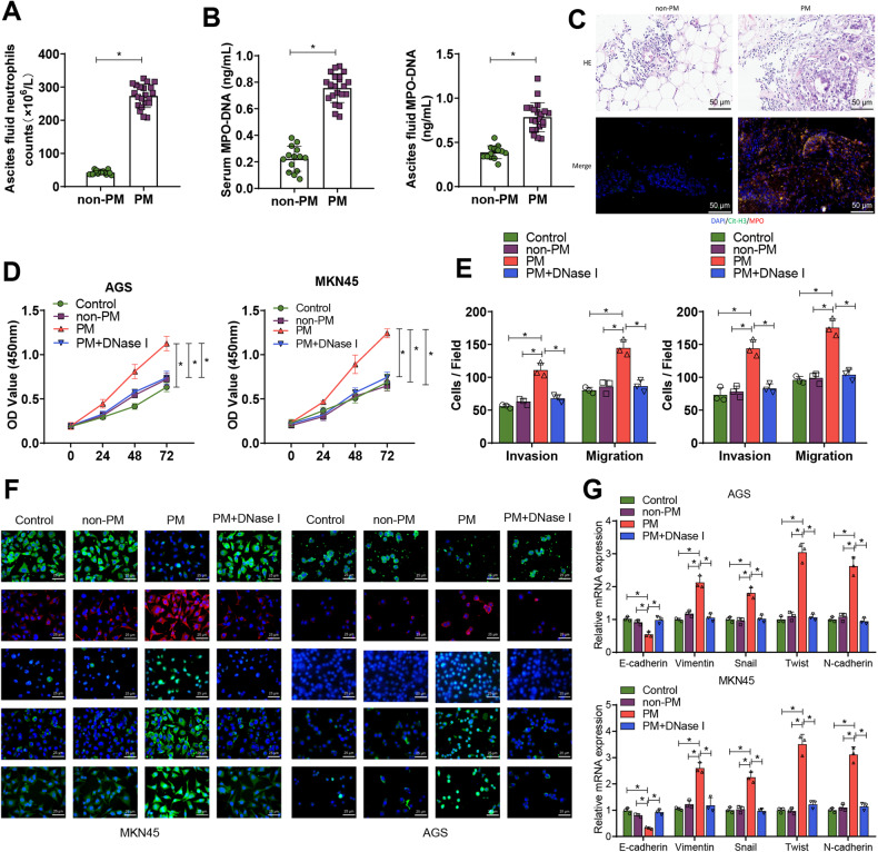
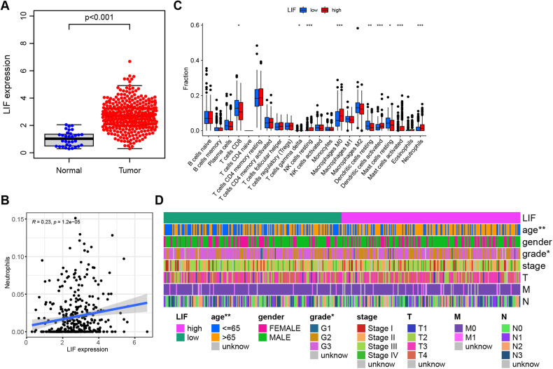
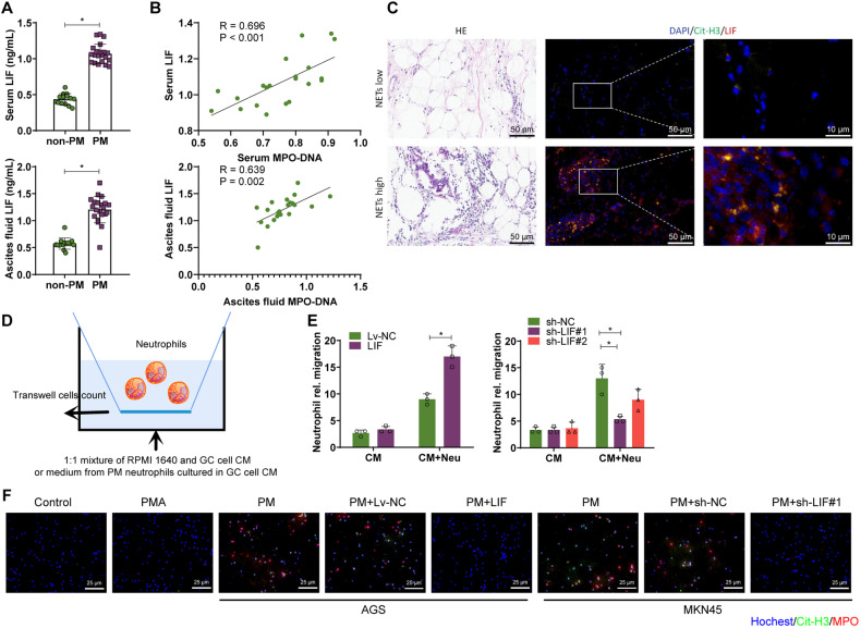
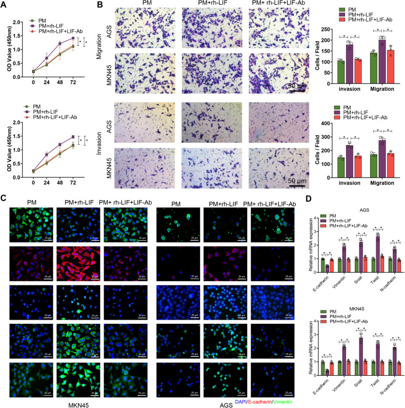
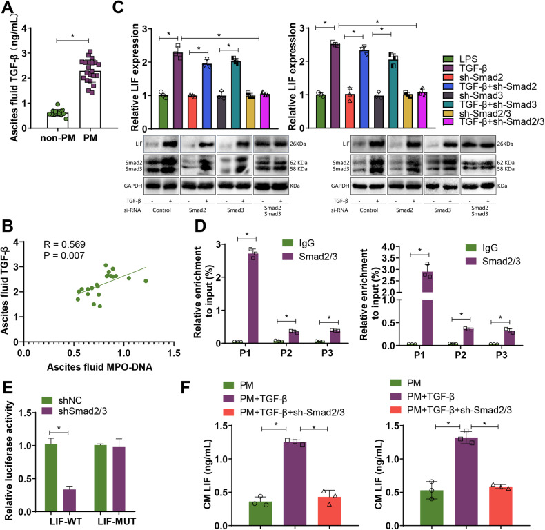
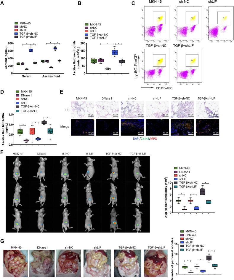
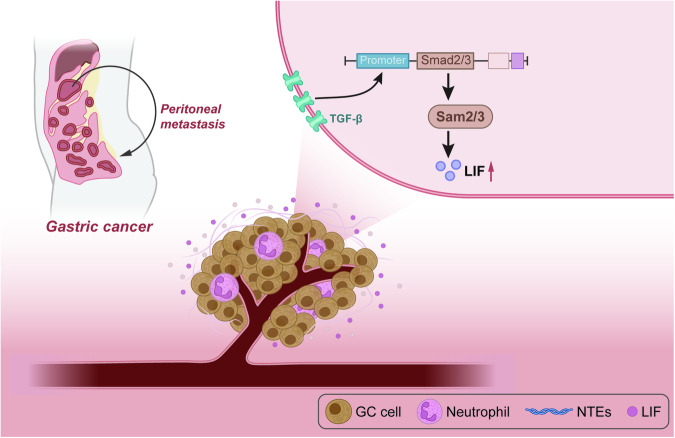
Gastric cancer (GC), notorious for its poor prognosis, often advances to peritoneal dissemination, a crucial determinant of detrimental outcomes. This study intricately explores the role of the TGFβ-Smad-LIF axis within the tumor microenvironment in propagating peritoneal metastasis, with a specific emphasis on its molecular mechanism in instigating Neutrophil Extracellular Traps (NETs) formation and encouraging GC cellular functions. Through a blend of bioinformatics analyses, utilizing TCGA and GEO databases, and meticulous in vivo and in vitro experiments, LIF was identified as pivotally associated with GC metastasis, notably, enhancing the NETs formation through neutrophil stimulation. Mechanistically, TGF-β was substantiated to elevate LIF expression via the activation of the Smad2/3 complex, culminating in NETs formation and consequently, propelling peritoneal metastasis of GC. This revelation uncovers a novel potential therapeutic target, promising a new avenue in managing GC and mitigating its metastatic propensities.


Conflict of interest statement

The authors declare no competing interests.

Figures

Fig. 1
Fig. 1. Effects of neutrophil NET formation on GC cell proliferation, invasion, migration, and EMT.
A Neutrophil counts in ascites were determined in the non-PM group (n = 12) and PM group (n = 18); B ELISA evaluated levels of MPO-DNA complexes in serum and ascites in the non-PM group (n = 12) and PM group (n = 18) to assess NETs levels; C Representative images of H&E and immunofluorescence staining of Cit-H3.MPO in omental tissues from non-PM group (n = 12) and PM group (n = 18), with blue indicating cell nuclei, green indicating Cit-H3, and red indicating MPO; D Proliferation ability of GC cells was detected by CCK-8 assay after co-culturing with neutrophils from each group; E Transwell assay was used to assess the migration and invasion ability of GC cells after co-culturing; F Immunofluorescence was used to detect the expression of EMT-related factors. E-cadherin and Snail were labeled in red, while Vimentin, Twist, and N-cadherin were labeled in green. The cell nuclei were labeled in blue. G RT-qPCR was performed to detect the expression of E-cadherin, Vimentin, Snail, Twist, and N-cadherin in GC cells after co-culture. *P < 0.05, cell experiments were repeated at least three times.
Fig. 2
Fig. 2. Key targets for tumor microenvironment neutrophil infiltration mediated by bioinformatics screening of GC peritoneal metastasis.
A Differential expression of LIF in the TCGA database: Normal, n = 37; Tumor, n = 375; B Correlation analysis of LIF and neutrophil infiltration, n = 375; C Analysis of differences in immune cell content between high expression group (red) and low expression group (green) of LIF; D Clinical correlation analysis of high and low expression of LIF. *P < 0.05, **P < 0.01, ***P < 0.001.
Fig. 3
Fig. 3. Influence of LIF on neutrophil recruitment and formation of NETs in the GC tumor microenvironment.
A Levels of LIF in serum and ascites of both non-PM group (n = 12) and PM group (n = 18) GC patients; B Pearson analysis of the correlation between serum MPO-DNA and LIF levels in PM group (n = 18) GC patients, and the correlation between ascites MPO-DNA and LIF levels; C Representative H&E and immunofluorescence co-staining images of Cit-H3 and LIF in the greater omentum tissue of PM group GC patients, with Hochest staining for nuclei in blue, Cit-H3 staining in green, and LIF staining in red; D Schematic diagram of the recruitment of neutrophils by CM from AGC cells or MKN-45 cells overexpressing or interfering with LIF, or by CM from neutral neutrophils (Neu) pretreated with CM from these cancer cells for 12 h; E Neutrophil counts in each group of migration; F Immunofluorescence co-staining detection of Cit-H3 and MPO levels, with Hochest staining for nuclei in blue, Cit-H3 staining in green, and MPO staining in red. *P < 0.05, cell experiments were repeated at least three times.
Fig. 4
Fig. 4. LIF activation affects GC cells’ proliferation, invasion, migration, and epithelial-mesenchymal transition (EMT) when co-cultured with neutrophils.
A After inducing co-cultivation of GC cells from PM with neutrophils, which were pretreated with 20 ng/ml recombinant LIF protein and 10 mg/mL neutralizing antibody against LIF, the proliferation ability of GC cells was assessed using the CCK-8 assay; B The migration and invasion ability of GC cells after co-cultivation was assessed using the Transwell assay; C Immunofluorescence staining to detect the expression of EMT-related factors. E-cadherin and Snail were labeled in red, while Vimentin, Twist, and N-cadherin were labeled in green. The cell nucleus was labeled in blue. D RT-qPCR to detect the expression of E-cadherin, Vimentin, Snail, Twist, and N-cadherin in GC cells after co-culture. *P < 0.05, cell experiments were repeated at least three times.
Fig. 5
Fig. 5. Transcriptional regulation of LIF expression by the TGF-β/Smad signaling axis.
A Levels of TGF-β in ascites of non-PM group (n = 12) and PM group (n = 18) GC patients; B Pearson analysis of the correlation between MPO-DNA in ascites of PM group (n = 18) GC patients and TGF-β levels; C Detection of LIF mRNA levels by RT-qPCR and protein expression of LIF, Smad2/3 by Western Blot; D ChIP-qPCR to detect potential binding sites of Smad2/3 in the LIF promoter region; E Dual-Luciferase reporter assay to investigate the transcriptional regulation of LIF by Smad2/3; F ELISA to measure the levels of LIF in GC cell culture medium. * Indicates that P < 0.05 and cell experiments are repeated at least three times.
Fig. 6
Fig. 6. The in vivo induction of PM by NETs may be associated with the activation of LIF.
A ELISA was used to measure the levels of LIF in serum and ascites of nude mice; B The neutrophil count in ascites of the PM nude mouse model was determined; C Flow cytometry was performed to analyze the proportion of neutrophils in ascites of nude mice; D ELISA was conducted to assess the levels of MPO-DNA complexes in the ascites of nude mice, indicating the levels of NETs; E Representative images of H&E and immunofluorescence staining of Cit-H3.MPO in the retinal tissue of nude mice, with cell nuclei labeled in blue, Cit-H3 in green, and MPO in red, scale bar = 50 μm; F Representative images and quantification of peritoneal metastatic lesions in PM nude mice. G Observation and statistical analysis of visible metastatic nodules on the mesentery of nude mice. N = 10.
Fig. 7
Fig. 7
Schematic diagram of the molecular mechanism by which the TGF-β-Smad-LIF axis in the tumor microenvironment promotes the formation of neutrophil extracellular traps (NETs) and facilitates peritoneal metastasis of gastric cancer.

References

    1. Smyth EC, Nilsson M, Grabsch HI, van Grieken NC, Lordick F. Gastric cancer. Lancet. 2020;396:635–48. doi: 10.1016/S0140-6736(20)31288-5. - DOI - PubMed
    1. Chia NY, Tan P. Molecular classification of gastric cancer. Ann Oncol. 2016;27:763–9. doi: 10.1093/annonc/mdw040. - DOI - PubMed
    1. Onoyama T, Ishikawa S, Isomoto H. Gastric cancer and genomics: review of literature. J Gastroenterol. 2022;57:505–16. doi: 10.1007/s00535-022-01879-3. - DOI - PMC - PubMed
    1. Sodek KL, Murphy KJ, Brown TJ, Ringuette MJ. Cell-cell and cell-matrix dynamics in intraperitoneal cancer metastasis. Cancer Metastasis Rev. 2012;31:397–414. doi: 10.1007/s10555-012-9351-2. - DOI - PMC - PubMed
    1. Rausei S, Ruspi L, Galli F, Pappalardo V, Di Rocco G, Martignoni F, et al. Seventh tumor-node-metastasis staging of gastric cancer: Five-year follow-up. World J Gastroenterol. 2016;22:7748–53. doi: 10.3748/wjg.v22.i34.7748. - DOI - PMC - PubMed

Publication types

MeSH terms

Substances