. 2024 Mar 15;14(1):6263.
doi: 10.1038/s41598-024-56936-z.

Role of gut-derived bacterial lipopolysaccharide and peripheral TLR4 in immobilization stress-induced itch aggravation in a mouse model of atopic dermatitis

Affiliations

Role of gut-derived bacterial lipopolysaccharide and peripheral TLR4 in immobilization stress-induced itch aggravation in a mouse model of atopic dermatitis

Da-Eun Cho et al. Sci Rep. .

Abstract

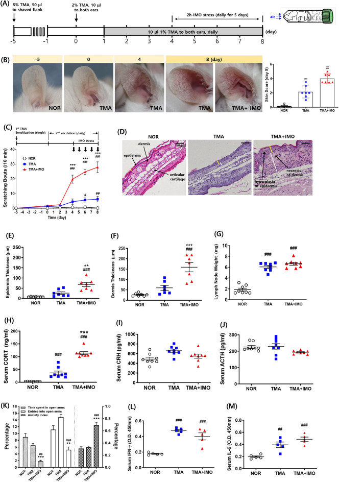
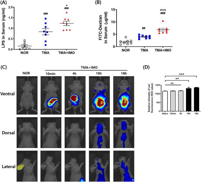
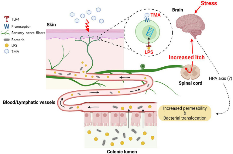
Psychological stress and intestinal leakage are key factors in atopic dermatitis (AD) recurrence and exacerbation. Here, we demonstrate the mechanism underlying bacterial translocation across intestinal epithelial barrier damaged due to stress and further aggravation of trimellitic anhydride (TMA)-induced itch, which remain unclear, in AD mice. Immobilization (IMO) stress exacerbated scratching bouts and colon histological damage, and increased serum corticosterone and lipopolysaccharide (LPS). Orally administered fluorescein isothiocyanate (FITC)-dextran and surgically injected (into the colon) Cy5.5-conjugated LPS were detected in the serum and skin after IMO stress, respectively. The relative abundance of aerobic or facultative anaerobic bacteria was increased in the colon mucus layer, and Lactobacillus murinus, E. coli, Staphylococcus nepalensis, and several strains of Bacillus sp. were isolated from the spleens and mesenteric lymph nodes. Oral antibiotics or intestinal permeability blockers, such as lubiprostone (Lu), 2,4,6-triaminopyrimidine (TAP) and ML-7, inhibited IMO stress-associated itch; however, it was reinduced through intradermal or i.p. injection of LPS without IMO stress. I.p. injection of TAK-242 (resatorvid), a TLR4 inhibitor, abrogated IMO stress-associated itch, which was also confirmed in TLR4-KO mice. IMO stress alone did not cause itch in naïve mice. IMO stress-induced itch aggravation in TMA-treated AD mice might be attributed to the translocation of gut-derived bacterial cells and LPS, which activates peripheral TLR4 signaling.

Keywords: Atopic dermatitis; Gut microbiota; Immobilization; Itch; Lipopolysaccharide; Stress; TLR4.


Conflict of interest statement

The authors declare no competing interests.

Figures

Figure 1
Figure 1
An experimental schedule of inducing AD and application of IMO stress (A), pictures of ear and skin, and skin score graph (B), and scratching bouts (C) in a mouse model of AD. (DF) Epidermis (E) and dermis (F) thickness based on hematoxylin and eosin-stained images (D), cervical lymph node weight (G), and serum levels of CORT (H), CRH (I), ACTH (J), IFN-γ (L), and IL-6 (M) were measured. EPM data are presented as relative percentages among groups (K). In EPM test, time spent in the open arms and open arm entries/total arm entries were recorded during a 5 min test session (n = 8 mice per group) in cohort 1. The anxiety index was calculated according to Cohen et al. as follows: Anxiety Index = 1-[([Open arm time/Test duration] + [Open arms entries/Total number of entries])/2]. At least three images of ear tissue sections per mouse were used to measure the epidermal and dermal thickness in E and F. The time points of IMO stress (a total of 5 instances) are denoted by the black arrows in C. Yellow and white bars indicate the thickness of the epidermis and dermis, respectively, in D. Scale bar = 50 μm in D. Error bars represent SEM #p < 0·05, ##p < 0·01, ###p < 0·001 versus NOR group; **p < 0·01, ***p < 0·001 versus TMA group. Two-way analysis of variance was used for EPM data. AD, atopic dermatitis; IMO, immobilization; CORT, corticosterone; CRH, corticotrophin-releasing hormone; ACTH, adrenocorticotropic hormone; IFN, interferon; IL, interleukin. TMA, trimellitic anhydride; NOR, normal; EPM, elevated plus maze.
Figure 2
Figure 2
Serum concentrations of LPS molecules (A) and FITC-dextran (B) after IMO stress in a mouse model of AD. In vivo fluorescence images (C) and a bar graph (D) showing the time-dependent distribution of Cy5.5-labeled bacterial LPS surgically injected into the colon after IMO stress in TMA-treated mice. Nude male mice on a BALB/c background were used for the in vivo immunofluorescence study. ##p < 0·01, ###p < 0·001 versus NOR group; *p < 0·05, ***p < 0·001 versus TMA group. LPS, lipopolysaccharides; FITC, fluorescein isothiocyanate; AD, atopic dermatitis; IMO, immobilization; TMA, trimellitic anhydride; NOR, normal.
Figure 3
Figure 3
Histological images (A) showing the H&E-stained intestinal epithelium and fluorescence-labeled LPS and occludin in paraffin-embedded distal colon sections. In H&E-stained images, blue lines indicate inner mucus layer length. Yellow arrows indicate cells immunopositive for LPS or occludin. Scratching bouts (B), the level of total bacteria in the feces (C), and serum levels of LPS molecules (D) and corticosterone (E) were assessed in TMA + IMO-treated mice under antibiotic cocktail (Abx) conditions. Black and white arrows indicate the time points of IMO stress (5 times) and Abx treatment (9 times), respectively. ns: not significant. **p < 0·01, ***p < 0·001 versus NOR group; p# < 0·05 versus TMA + IMO group. IM: inner mucus layer; LPS, lipopolysaccharides; IMO, immobilization; TMA, trimellitic anhydride; NOR, normal; hematoxylin and eosin.
Figure 4
Figure 4
The relative abundance of strict aerobes and facultative anaerobes increased more (or only detected) in the mucus layers of TMA + IMO mice than in NOR mice due to IMO stress (A) and in the mucus layers of NOR mice than in TMA + IMO mice (B). Red and blue bars indicate bacterial genera that overgrew in NOR and TMA + IMO mice, respectively (n = 3 in each group). Each bar indicates an average value of three mice and is expressed as the mean ± standard error of the mean (SEM). TMA: trimellitic anhydride, IMO: immobilization; NOR, normal.
Figure 5
Figure 5
The effects of i.d. and i.p. injections of LPS molecules on the scratching behavior (A, D) and epidermis and dermis thickness of ear tissue (B, E), respectively, in TMA-treated mice. I.d.-injected LPS molecules were immunostained in the TMA + LPS_20 ng/ear i.d. group on day 8 (C). The serum concentration of i.p. injected LPS molecules was measured in the TMA + LPS_20 μg/mouse i.p. group on day 12 (F). Black arrows in A and D indicate the time points of LPS injections. The lower figures in both the NOR and LPS groups depict an enlargement of the orange squares seen in the upper figures. *p < 0·05, **p < 0·01, ***p < 0·001 versus TMA + saline i.d. (or i.p.) group. LPS, lipopolysaccharides; TMA, trimellitic anhydride; NOR, normal; i.d., intradermal; i.p., intraperitoneal.
Figure 6
Figure 6
Effects of lubiprostone (Lu), ML-7, TAP, and TAK-242 on scratching behavior (A, B, C, G), epidermis, and dermis thickness of ear tissue (D, E, F, J), respectively, in the TMA + IMO-treated group. Lu was administered at two different doses: 100 μg/kg in the TMA + IMO + Lu-h group and 20 μg/kg in the TMA + IMO + Lu-l group. White arrows indicate the time points of inhibitor treatments in A, B, C, and D. Scratching bouts (H) and serum levels of LPS (K) and corticosterone (L) were assessed in TLR4 KO male mice on a C57BL/6 background with TMA or TMA + IMO treatment. The time points of IMO stress (a total of 5 instances) are denoted by the black arrows in E. In the small box in H, scratching behaviors of TMA-and TMA + IMO-treated naïve mice of the C57BL/6 background are presented as a control. TLR4 protein (yellow-stained) in ear tissues was immunostained with PGP9.5 (red-stained, a marker for peripheral nerve fibers) in the TMA + IMO-treated group on day 8 (I). White arrows indicate representative immunostained spots of TLR4 proteins in the TMA- and TMA + IMO-treated groups. The border line between the epidermis and dermis layers is indicated by the yellow lines in F. *p < 0·05, **p < 0·01, ***p < 0·001 versus TMA group (or TMA_KO); $$$p < 0·001 versus TMA + IMO-treated group. TMA, trimellitic anhydride; NOR, normal; KO, knockout; TLR4, Toll□like receptor 4; IMO, immobilization.
Figure 7
Figure 7
A proposed mechanism of translocation of gut-derived bacterial LPS molecules across intestinal epithelium and the peripheral activation of TLR4 in IMO stress-induced itch aggravation in a mouse model of TMA-induced AD (created with BioRender.com).

Similar articles

Cited by

References

    1. Kodama A, Horikawa T, Suzuki T, Ajiki W, Takashima T, Harada S, Ichihashi M. Effect of stress on atopic dermatitis: Investigation in patients after the great hanshin earthquake. J. Allergy Clin. Immunol. 1999;104:173–176. doi: 10.1016/S0091-6749(99)70130-2. - DOI - PubMed
    1. Picardi A, Abeni D. Stressful life events and skin diseases: Disentangling evidence from myth. Psychother. Psychosom. 2001;70:118–136. doi: 10.1159/000056237. - DOI - PubMed
    1. Kilpeläinen M, Koskenvuo M, Helenius H, Terho EO. Stressful life events promote the manifestation of asthma and atopic diseases. Clin. Exp. Allergy. 2000;32:256–263. doi: 10.1046/j.1365-2222.2002.01282.x. - DOI - PubMed
    1. Kelsay K, Klinnert M, Bender B. Addressing psychosocial aspects of atopic dermatitis. Immunol. Allergy Clin. N. Am. 2010;30:385–396. doi: 10.1016/j.iac.2010.05.003. - DOI - PubMed
    1. Hanning N, Edwinson AL, Ceuleers H, Peters SA, De Man JG, Hassett LC, De Winter BY, Grover M, et al. Intestinal barrier dysfunction in irritable bowel syndrome: A systematic review. Ther. Adv. Gastroenterol. 2021;14:1–31. doi: 10.1177/1756284821993586. - DOI - PMC - PubMed