Tetrahedral framework nucleic acids for improving wound healing
- PMID: 38491372
- PMCID: PMC10943864
- DOI: 10.1186/s12951-024-02365-z
Tetrahedral framework nucleic acids for improving wound healing
Abstract
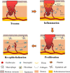
Wounds are one of the most common health issues, and the cost of wound care and healing has continued to increase over the past decade. In recent years, there has been growing interest in developing innovative strategies to enhance the efficacy of wound healing. Tetrahedral framework nucleic acids (tFNAs) have emerged as a promising tool for wound healing applications due to their unique structural and functional properties. Therefore, it is of great significance to summarize the applications of tFNAs for wound healing. This review article provides a comprehensive overview of the potential of tFNAs as a novel therapeutic approach for wound healing. In this review, we discuss the possible mechanisms of tFNAs in wound healing and highlight the role of tFNAs in modulating key processes involved in wound healing, such as cell proliferation and migration, angiogenesis, and tissue regeneration. The targeted delivery and controlled release capabilities of tFNAs offer advantages in terms of localized and sustained delivery of therapeutic agents to the wound site. In addition, the latest research progress on tFNAs in wound healing is systematically introduced. We also discuss the biocompatibility and biosafety of tFNAs, along with their potential applications and future directions for research. Finally, the current challenges and prospects of tFNAs are briefly discussed to promote wider applications.
Keywords: DNA nanomaterials; Tetrahedral framework nucleic acids; Tissue regeneration; Wound healing.
© 2024. The Author(s).
Conflict of interest statement
The authors declare no competing interests.
Figures


Similar articles
-
Tetrahedral framework nucleic acids promote diabetic wound healing via the Wnt signalling pathway.Cell Prolif. 2022 Nov;55(11):e13316. doi: 10.1111/cpr.13316. Epub 2022 Jul 22. Cell Prolif. 2022. PMID: 35869570 Free PMC article.
-
Tetrahedral Framework Nucleic Acids Promote Corneal Epithelial Wound Healing in Vitro and in Vivo.Small. 2019 Aug;15(31):e1901907. doi: 10.1002/smll.201901907. Epub 2019 Jun 13. Small. 2019. PMID: 31192537
-
Antioxidative and Angiogenesis-Promoting Effects of Tetrahedral Framework Nucleic Acids in Diabetic Wound Healing with Activation of the Akt/Nrf2/HO-1 Pathway.ACS Appl Mater Interfaces. 2020 Mar 11;12(10):11397-11408. doi: 10.1021/acsami.0c00874. Epub 2020 Feb 28. ACS Appl Mater Interfaces. 2020. PMID: 32083455
-
Recent progress and application of the tetrahedral framework nucleic acid materials on drug delivery.Expert Opin Drug Deliv. 2023 Jul-Dec;20(11):1511-1530. doi: 10.1080/17425247.2023.2276285. Epub 2023 Dec 20. Expert Opin Drug Deliv. 2023. PMID: 37898874 Review.
-
Functionalizing tetrahedral framework nucleic acids-based nanostructures for tumor in situ imaging and treatment.Colloids Surf B Biointerfaces. 2024 Aug;240:113982. doi: 10.1016/j.colsurfb.2024.113982. Epub 2024 May 21. Colloids Surf B Biointerfaces. 2024. PMID: 38788473 Review.
Cited by
-
Targeted siRNA Delivery Against RUNX1 Via tFNA: Inhibiting Retinal Neovascularization and Restoring Vessels Through Dll4/Notch1 Signaling.Invest Ophthalmol Vis Sci. 2025 Mar 3;66(3):39. doi: 10.1167/iovs.66.3.39. Invest Ophthalmol Vis Sci. 2025. PMID: 40105819 Free PMC article.
-
Advances and Challenges in Immune-Modulatory Biomaterials for Wound Healing Applications.Pharmaceutics. 2024 Jul 26;16(8):990. doi: 10.3390/pharmaceutics16080990. Pharmaceutics. 2024. PMID: 39204335 Free PMC article. Review.
-
Ultrafast enzyme-responsive hydrogel for real-time assessment and treatment optimization in infected wounds.J Nanobiotechnology. 2025 Jan 8;23(1):9. doi: 10.1186/s12951-024-03078-z. J Nanobiotechnology. 2025. PMID: 39780182 Free PMC article.
-
Biomaterial-Based Nucleic Acid Delivery Systems for In Situ Tissue Engineering and Regenerative Medicine.Int J Mol Sci. 2025 Jul 30;26(15):7384. doi: 10.3390/ijms26157384. Int J Mol Sci. 2025. PMID: 40806513 Free PMC article. Review.
-
Update on the Study of Angiogenesis in Surgical Wounds in Patients with Childhood Obesity.Biomedicines. 2025 Feb 5;13(2):375. doi: 10.3390/biomedicines13020375. Biomedicines. 2025. PMID: 40002788 Free PMC article. Review.
References
-
- Wang X, Chang J, Wu C. Bioactive inorganic/organic nanocomposites for wound healing. Appl Mater Today. 2018;11:308–19. doi: 10.1016/j.apmt.2018.03.001. - DOI
-
- Zhao X, Pei D, Yang Y, Xu K, Yu J, Zhang Y, Zhang Q, He G, Zhang Y, Li A, et al. Green Tea Derivative Driven Smart Hydrogels with Desired functions for Chronic Diabetic Wound Treatment. Adv Funct Mater. 2021;31:2009442. doi: 10.1002/adfm.202009442. - DOI
Publication types
MeSH terms
Substances
Grants and funding
LinkOut - more resources
Full Text Sources
Miscellaneous

