. 2024 Mar 15;24(1):130.
doi: 10.1186/s12890-024-02940-y.

IRF4-mediated Treg phenotype switching can aggravate hyperoxia-induced alveolar epithelial cell injury

Affiliations

IRF4-mediated Treg phenotype switching can aggravate hyperoxia-induced alveolar epithelial cell injury

He Langyue et al. BMC Pulm Med. .

Abstract

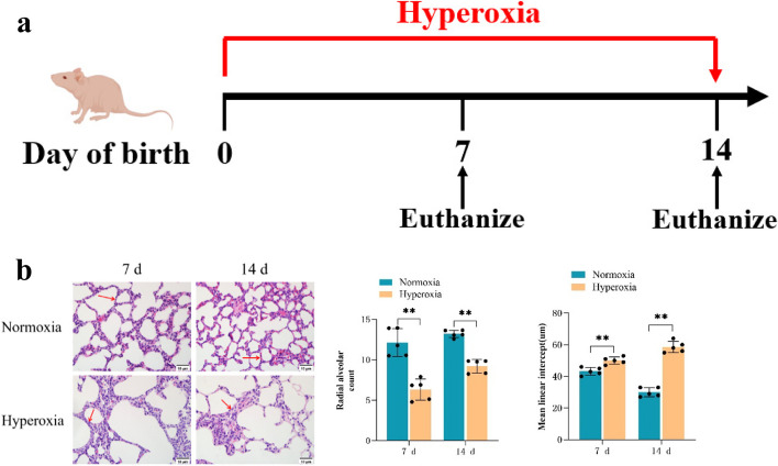
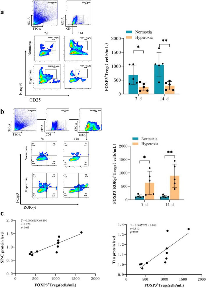
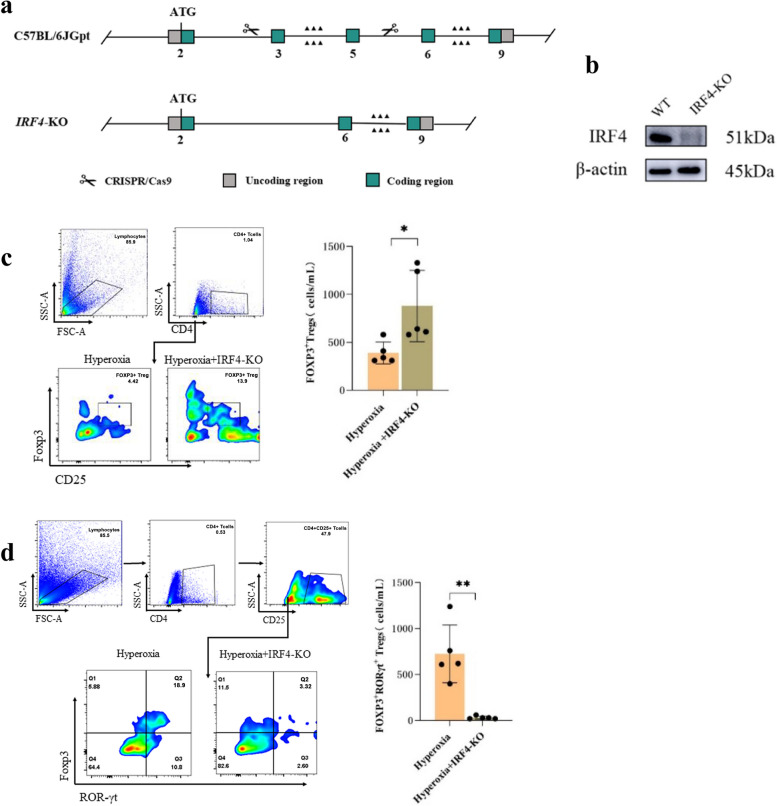
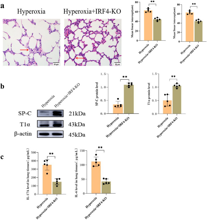
Bronchopulmonary dysplasia (BPD) is characterized by alveolar dysplasia, and evidence indicates that interferon regulatory factor 4 (IRF4) is involved in the pathogenesis of various inflammatory lung diseases. Nonetheless, the significance and mechanism of IRF4 in BPD remain unelucidated. Consequently, we established a mouse model of BPD through hyperoxia exposure, and ELISA was employed to measure interleukin-17 A (IL-17 A) and interleukin-6 (IL-6) expression levels in lung tissues. Western blotting was adopted to determine the expression of IRF4, surfactant protein C (SP-C), and podoplanin (T1α) in lung tissues. Flow cytometry was utilized for analyzing the percentages of FOXP3+ regulatory T cells (Tregs) and FOXP3+RORγt+ Tregs in CD4+ T cells in lung tissues to clarify the underlying mechanism. Our findings revealed that BPD mice exhibited disordered lung tissue structure, elevated IRF4 expression, decreased SP-C and T1α expression, increased IL-17 A and IL-6 levels, reduced proportion of FOXP3+ Tregs, and increased proportion of FOXP3+RORγt+ Tregs. For the purpose of further elucidating the effect of IRF4 on Treg phenotype switching induced by hyperoxia in lung tissues, we exposed neonatal mice with IRF4 knockout to hyperoxia. These mice exhibited regular lung tissue structure, increased proportion of FOXP3+ Tregs, reduced proportion of FOXP3+RORγt+ Tregs, elevated SP-C and T1α expression, and decreased IL-17 A and IL-6 levels. In conclusion, our findings demonstrate that IRF4-mediated Treg phenotype switching in lung tissues exacerbates alveolar epithelial cell injury under hyperoxia exposure.

Keywords: Bronchopulmonary dysplasia; Forkhead transcription factor 3; Interferon regulatory factor 4; Regulatory T cells; Retinoic acid-related orphan nuclear receptor.


Conflict of interest statement

The authors declare no competing interests.

Figures

Fig. 1
Fig. 1
Analysis of lung histopathology and alveolar count in mice. a C57BL/6 mice were exposed to hyperoxia at birth and were sacrificed on days 7 and 14. b HE staining of lung tissues from mice in the air and hyperoxia groups (Scale bar = 10 μm; 400×), showed a signifcant decrease in radial alveolar count, and a signifcant increase in mean linear intercept. The representative data from five independent experiments. Data are presented as mean ± SD (n = 5, t-test one-way); *P < 0.05 and **P < 0.01, vs. normoxia group
Fig. 2
Fig. 2
Abnormal transdifferentiation and inflammatory response induced by hyperoxia in mice. a Western blot analysis of SP-C, T1α, and IRF4 expression in the mouse lung tissues of the normoxia and hyperoxia groups. β-actin was used as the loading control. b ELISA of IL-6 and IL-17 A expression in the mouse lung tissues of each group. c Correlation analysis of IRF4 with IL-6 and IL-17 A. The representative data from five independent experiments. Data are presented as mean ± SD (n = 5, t-test one-way); *P < 0.05 and **P < 0.01 vs. normoxia group
Fig. 3
Fig. 3
Hyperoxia-induced conversion of FOXP3 + Tregs to FOXP3 + RORγt + Tregs in mouse lung tissues. a Flow cytometry analysis of FOXP+ Tregs in the mouse lung tissues of the normoxia and hyperoxia groups. b Flow cytometry analysis of FOXP3+RORγt+ Tregs in the mouse lung tissues of the normoxia and hyperoxia groups. c Correlation analysis of FOXP3+ Tregs with SP-C and T1α. The representative data from five independent experiments. Data are presented as mean ± SD (n = 5, t-test one-way); *P < 0.05 and **P < 0.01 vs. normoxia group
Fig. 4
Fig. 4
IRF4 knockdown attenuates the conversion of FOXP3+ Tregs to FOXP3+RORγt+ Tregs in the lung tissues of mice after hyperoxia induction. a Schematic diagram of the construction of IRF4-KO mice by CRISPR/Cas9 technology. b Deletion of IRF4 was confirmed by Western blotting (n = 5). c Flow cytometry analysis of FOXP3+ Tregs in the mouse lung tissues of WT-hyperoxia and KO-hyperoxia groups. d Flow cytometry analysis of FOXP3+RORγt+ Tregs in the mouse lung tissues of the WT-hyperoxia and KO-hyperoxia groups. The representative data from five independent experiments. Data are presented as mean ± SD (n = 5, t-test one-way); *P < 0.05 and **P < 0.01 vs. WT-hyperoxia group
Fig. 5
Fig. 5
Improvement of hyperoxia-induced lung tissue injury by IRF4 knockout. a HE staining of lung tissues from mice in the WT-hyperoxia and KO-hyperoxia groups (Scale bar = 10 μm; 400×), showed a significant increase in radial alveolar count, and a significant decrease in mean linear intercept. b Western blot analysis of SP-C and T1α protein expression levels in the lung tissues of WT-hyperoxia and KO-hyperoxia groups. c ELISA of IL-6 and IL-17 A expression in the mouse lung tissues of WT-hyperoxia and KO-hyperoxia groups. The representative data from five independent experiments. Data are presented as mean ± SD (n = 5, t-test one-way); *P < 0.05 and **P < 0.01 vs. WT-hyperoxia
Fig. 6
Fig. 6
Graphical Abstract. Mechanism of IRF4 regulation of hyperoxia-induced lung injury through conversion of FOXP3+ Tregs to FOXP3+RORγt+ Tregs under hyperoxia exposure

References

    1. Moschino L, Bonadies L, Baraldi E. Lung growth and pulmonary function after prematurity and bronchopulmonary dysplasia[J] Pediatr Pulmonol. 2021;56(11):3499–508. doi: 10.1002/ppul.25380. - DOI - PMC - PubMed
    1. Liberti DC, Kremp MM, Liberti WA, et al. Alveolar epithelial cell fate is maintained in a spatially restricted manner to promote lung regeneration after acute injury[J] Cell Rep. 2021;35(6):109092. doi: 10.1016/j.celrep.2021.109092. - DOI - PMC - PubMed
    1. Deng X, Bao Z, Yang X, et al. Molecular mechanisms of cell death in bronchopulmonary dysplasia[J] Apoptosis: Int J Program Cell Death. 2023;28(1–2):39–54. doi: 10.1007/s10495-022-01791-4. - DOI - PubMed
    1. Tao Y, Han X, Guo W-L. Predictors of Bronchopulmonary Dysplasia in 625 neonates with respiratory distress Syndrome[J] J Trop Pediatr. 2022;68(3):fmac037. doi: 10.1093/tropej/fmac037. - DOI - PMC - PubMed
    1. Hirani D, Alvira CM, Danopoulos S, et al. Macrophage-derived IL-6 trans-signalling as a novel target in the pathogenesis of bronchopulmonary dysplasia[J] Eur Respir J. 2022;59(2):2002248. doi: 10.1183/13993003.02248-2020. - DOI - PMC - PubMed

MeSH terms

Substances