. 2024 Mar 27;146(12):8058-8070.
doi: 10.1021/jacs.3c12037. Epub 2024 Mar 16.

A Compact Reprogrammed Genetic Code for De Novo Discovery of Proteolytically Stable Thiopeptides

Affiliations

A Compact Reprogrammed Genetic Code for De Novo Discovery of Proteolytically Stable Thiopeptides

Alexander A Vinogradov et al. J Am Chem Soc. .

Abstract

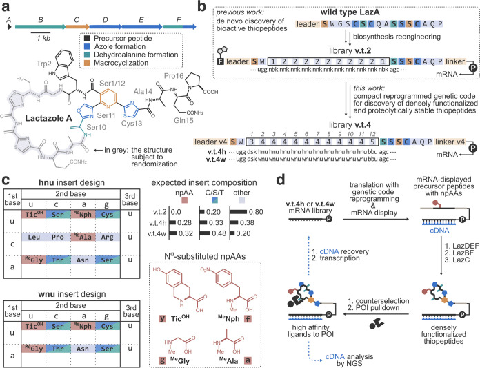
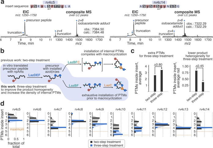
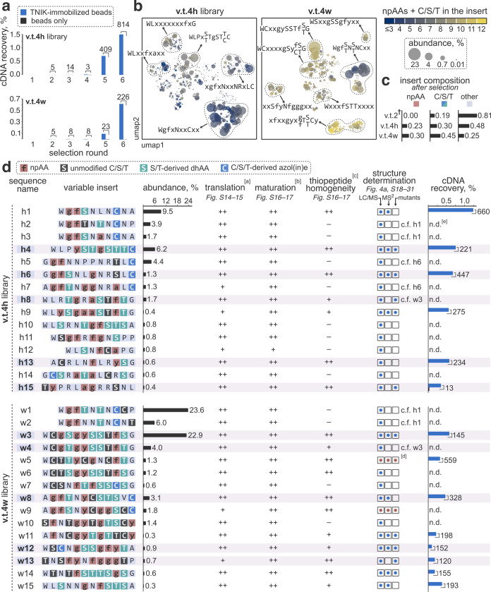
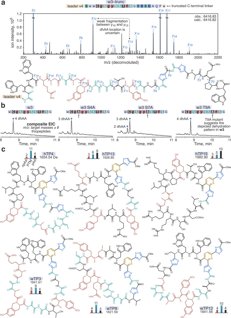
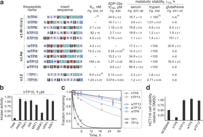
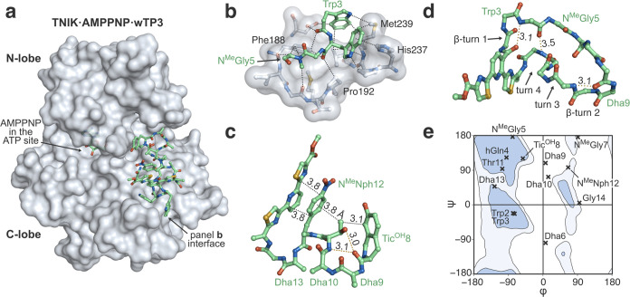
Thiopeptides make up a group of structurally complex peptidic natural products holding promise in bioengineering applications. The previously established thiopeptide/mRNA display platform enables de novo discovery of natural product-like thiopeptides with designed bioactivities. However, in contrast to natural thiopeptides, the discovered structures are composed predominantly of proteinogenic amino acids, which results in low metabolic stability in many cases. Here, we redevelop the platform and demonstrate that the utilization of compact reprogrammed genetic codes in mRNA display libraries can lead to the discovery of thiopeptides predominantly composed of nonproteinogenic structural elements. We demonstrate the feasibility of our designs by conducting affinity selections against Traf2- and NCK-interacting kinase (TNIK). The experiment identified a series of thiopeptides with high affinity to the target protein (the best KD = 2.1 nM) and kinase inhibitory activity (the best IC50 = 0.15 μM). The discovered compounds, which bore as many as 15 nonproteinogenic amino acids in an 18-residue macrocycle, demonstrated high metabolic stability in human serum with a half-life of up to 99 h. An X-ray cocrystal structure of TNIK in complex with a discovered thiopeptide revealed how nonproteinogenic building blocks facilitate the target engagement and orchestrate the folding of the thiopeptide into a noncanonical conformation. Altogether, the established platform takes a step toward the discovery of thiopeptides with high metabolic stability for early drug discovery applications.


Conflict of interest statement

The authors declare the following competing financial interest(s): A.V., Y.Z., Y.G. and H.S. are listed as co-inventors on the patents pertaining to lactazole engineering. Other authors declare no competing interests.

Figures

Figure 1
Figure 1
Design of the thiopeptide/mRNA display platform with a compact reprogrammed genetic code. (a) Biosynthetic gene cluster and the chemical structure of lactazole A, a thiopeptide whose reengineered biosynthesis serves as the foundation for the discovery platform. (b) Reengineering of lactazole biosynthesis. Previous work reported the construction of mRNA display library v.t.2. In this work, the libraries are redesigned to accommodate compact reprogrammed genetic codes for the discovery of densely functionalized thiopeptides. See also Figure S2 for the complete description of v.t.4 library designs. (c) Reprogrammed genetic code tables for translation. Library v.t.4h and v.t.4w contain random inserts composed of hnu- and wnu-encoded amino acids, respectively. These degenerate codons, particularly wnu, lead to a high density of C/S/T residues and npAAs in the thiopeptide inserts compared to the previously reported library v.t.2. (d) Selection scheme. mRNA-barcoded precursor peptides are produced using the constructed translation system, and upon sequential three-step treatment with LazDEF/LazBF/LazC are converted to thiopeptides. The resulting library is subjected to a pulldown against immobilized TNIK to enrich for high-affinity protein ligands. The process is repeated by recovering cDNA, amplifying it by PCR, and transcribing it to mRNA for the following round of selection. POI: protein of interest.
Figure 2
Figure 2
Construction of the selection platform. (a) Translational incorporation of multiple npAAs into partially randomized LazA variants. Peptides rv4c5 and rv4c14 were expressed with the FIT system using the reprogrammed genetic code (S. I. 2.6), and the translation outcomes were analyzed by LC/MS. Displayed are extracted ion current (EIC) chromatograms and composite MS spectra integrated over peptide-derived peaks. Formation of the expected peptides as the major product was observed in both cases. The left arrows shown below the insert sequences indicate the location of the truncations. *: translation protein carryover. (b) Development of a three-step thiopeptide maturation protocol, in which LazA-derived precursor peptides are sequentially treated with LazDEF/LazBF/LazC. (c) The use of the three-step protocol improves both the density of PTMs (average number of PTMs inside insert) and the homogeneity of the resulting products (average relative standard deviation of the number of PTMs inside insert) compared to the previously employed two-step treatment. The statistics are derived from the analysis of the rv4c5–14 maturation products by LC/MS ((d) and Figures S4–S13).
Figure 3
Figure 3
Results of the affinity selections against TNIK. (a) The progress of the selections as monitored by the cDNA recovery rate. Numbers above the bars indicate the ratio of cDNA recovery between TNIK pulldown and counterselection fractions. (b) Uniform manifold approximation and projection (umap) embedding of top 1000 most abundant random insert sequences obtained after six rounds of selection. The sequences are colored according to the number of npAAs and C/S/T residues in the insert. (c) Insert compositions in round six libraries. As designed, libraries v.t.4h and v.t.4w furnished sequences with high npAA/C/S/T content. : Data for the previously reported v.t.2 selection is provided for comparison. (d) Initial characterization of the selected hits. Shown are the random insert sequences with the established modification pattern. Here and elsewhere in the text, residues highlighted in black represent unmodified C/S/T residues; in red—translationally installed npAAs (Figure 1c for chemical structures and one-letter codes); in blue—C/S/T-derived azol(in)es; in green—S/T-derived dhAAs (Dha and Dhb). The sequences selected for resynthesis are highlighted in light pink. [a]: scale: ++/+/–, where “++” indicates expected peptide as the major translation product and “+” indicates expected peptide accompanied by major side products. [b]: the efficiency of the enzymatic maturation; scale: ++/+/–, where “++” indicates the full conversion to thiopeptides with no/minimal accumulation of partially modified peptides and shunt products. [c]: scale: ++/+/–, with “++” and “+” indicating the cases where a single species accounted for ≥85% and ≥70% of the observed thiopeptide products, respectively. [d]: for w5 and w9, reliable structures could not be ascertained. [e]: n.d.: not determined.
Figure 4
Figure 4
Elucidation of the structures of the discovered hits using w3 as an example. (a) MS/MS analysis of the insert modification pattern in C-terminally truncated w3 precursor peptide (S. I. 2.6–2.7). Shown is a zoomed-in section of a charge-deconvoluted CID fragmentation spectrum for the LazDEF/LazBF-modified w3–trunc; b-ion assignments and neutral molecule losses are omitted for clarity. Fragmentation assignments are mapped onto the suggested chemical structure of the modified w3–trunc shown below. (b) Mutational analysis results. The specified w3 mutants were expressed with the reprogrammed translation system and sequentially treated with LazDEF/LazBF/LazC. Reaction outcomes were analyzed by LC/MS. Displayed are composite EIC chromatograms for the detected thiopeptide products (scaled Y-axes). The combination of MS/MS and mutational analyses enabled an unambiguous structural assignment for wTP3. (c) Example chemical structures of the identified hit thiopeptides. The top and bottom rows feature the structures derived from library v.t.4h and v.t.4w, respectively. The bar graphs denote the numbers of npAA, PTM, and proteinogenic amino acids (pAAs) in the structures.
Figure 5
Figure 5
Biochemical characterization of the synthesized thiopeptides. (a) Values for binding affinity to TNIK (KD; average from three multicycle kinetic experiments; data fit assuming the 1:1 binding model; see also Table S5); in vitro kinase inhibition (IC50 against TNIK derived from at least two ADP-Glo-based experiments conducted in triplicate each; data fit to the standard 4-parameter logistic curve); and metabolic stability in the presence of human serum, trypsin, and glutathione (half-life, τ1/2; a single experiment conducted in triplicate; data fit to the standard first-order exponential decay curve). [a]: no measurable binding/inhibition [b]: binding and inhibition data for TP1 and TP15 are taken from ref (33). [c]: the compound did not completely inhibit the kinase even at high concentrations (see Figure S35) [d]: no measurable degradation during the course of the experiment [e]: not determined. (b) Kinase selectivity profiling outcomes for hTP15, tested at 1 μM thiopeptide concentration. Analogous data for hTP8 is summarized in Figure S36. (c) Degradation of the five best compounds (hTP8, hTP13, wTP8, wTP12, and wTP13) in human serum compared to the previously discovered TP1 and TP15. (d) Cellular activity of the discovered thiopeptides in HCT116 cells. Shown are the cell viability results after a 24 h incubation with the compounds (S.I. 2.11). Thiopeptides hTP8 and wTP12 as well as the positive control (NCB0846) were assayed at 10 μM concentration; hTP15 and wTP3, at 8 μM.
Figure 6
Figure 6
Structural analysis of the interaction between TNIK and wTP3. (a) Overview of the X-ray crystal structure of the TNIK·AMPPNP·wTP3 complex (pdb 8wm0). The protein surface is shown in gray; wTP3 and AMPPNP are displayed as ball and stick models. (b) Hydrophobic interactions between αG-helix and P+1 loop of TNIK and wTP3. Atoms within 4 Å distance are connected with dotted lines. (c) Intramolecular interactions in wTP3 with key distances highlighted. Hydrogen bonds are shown as yellow dotted lines. (d) Folding of TNIK-bound wTP3. The protein and amino acid side chains are omitted for clarity. The thiopeptide folds into a noncanonical structure featuring four turns. (e) Plot of φ/ψ dihedral angles of TNIK-bound wTP3. Inner and outer contours enclose 98 and 99.5% of the structures in Protein Data Bank, respectively. wTP3 contains several amino acids outside of the canonical Ramachandran space.

Similar articles

Cited by

References

    1. Montalbán-López M.; Scott T. A.; Ramesh S.; Rahman I. R.; van Heel A. J.; Viel J. H.; Bandarian V.; Dittmann E.; Genilloud O.; Goto Y.; Grande Burgos M. J.; Hill C.; Kim S.; Koehnke J.; Latham J. A.; Link A. J.; Martínez B.; Nair S. K.; Nicolet Y.; Rebuffat S.; Sahl H.-G.; Sareen D.; Schmidt E. W.; Schmitt L.; Severinov K.; Süssmuth R. D.; Truman A. W.; Wang H.; Weng J.-K.; van Wezel G. P.; Zhang Q.; Zhong J.; Piel J.; Mitchell D. A.; Kuipers O. P.; van der Donk W. A. New Developments in RiPP Discovery, Enzymology and Engineering. Nat. Prod. Rep. 2021, 38, 130–239. 10.1039/D0NP00027B. - DOI - PMC - PubMed
    1. Arnison P. G.; Bibb M. J.; Bierbaum G.; Bowers A. A.; Bugni T. S.; Bulaj G.; Camarero J. A.; Campopiano D. J.; Challis G. L.; Clardy J.; Cotter P. D.; Craik D. J.; Dawson M.; Dittmann E.; Donadio S.; Dorrestein P. C.; Entian K.; Gruber C. W.; Fischbach M. A.; Garavelli J. S.; Ulf G.; Haft D. H.; Hemscheidt T. K.; Hertweck C.; Hill C.; Horswill A. R.; Jaspars M.; Kelly W. L.; Klinman J. P.; Kuipers O. P.; Link A. J.; Liu W.; Marahiel M. A.; Nair S. K.; Mitchell D. A.; Moll G. N.; Moore B. S.; Rolf M.; Nes I. F.; Norris G. E.; Olivera B. M.; Onaka H.; Patchett M. L.; Piel J.; Reaney M. J. T.; Ross R. P.; Sahl H.; Schmidt E. W.; Selsted M. E.; Severinov K.; Shen B.; Sivonen K.; Smith L.; Stein T.; Tagg J. R.; Tang G.; Truman A. W.; Vederas J. C.; Walsh C. T.; Walton J. D.; Wenzel S. C.; Willey J. M.; van der Donk W. A.; et al. Ribosomally Synthesized and Post-Translationally Modified Peptide Natural Products: Overview and Recommendations for a Universal Nomenclature. Nat. Prod. Rep. 2013, 30, 108–160. 10.1039/C2NP20085F. - DOI - PMC - PubMed
    1. Bagley M. C.; Dale J. W.; Merritt E. A.; Xiong X. Thiopeptide Antibiotics. Chem. Rev. 2005, 105, 685–714. 10.1021/cr0300441. - DOI - PubMed
    1. Harms J. M.; Wilson D. N.; Schluenzen F.; Connell S. R.; Stachelhaus T.; Zaborowska Z.; Spahn C. M. T.; Fucini P. Translational Regulation via L11: Molecular Switches on the Ribosome Turned On and Off by Thiostrepton and Micrococcin. Mol. Cell 2008, 30, 26–38. 10.1016/j.molcel.2008.01.009. - DOI - PubMed
    1. Cundliffe E.; Thompson J. Concerning the Mode of Action of Micrococcin upon Bacterial Protein Synthesis. Eur. J. Biochem. 1981, 118, 47–52. 10.1111/j.1432-1033.1981.tb05484.x. - DOI - PubMed

Publication types

LinkOut - more resources