. 2024 Mar 16;24(1):53.
doi: 10.1007/s10238-024-01311-5.

Use of tryptic peptide MALDI mass spectrometry imaging to identify the spatial proteomic landscape of colorectal cancer liver metastases

Affiliations

Use of tryptic peptide MALDI mass spectrometry imaging to identify the spatial proteomic landscape of colorectal cancer liver metastases

Celine Man Ying Li et al. Clin Exp Med. .

Abstract

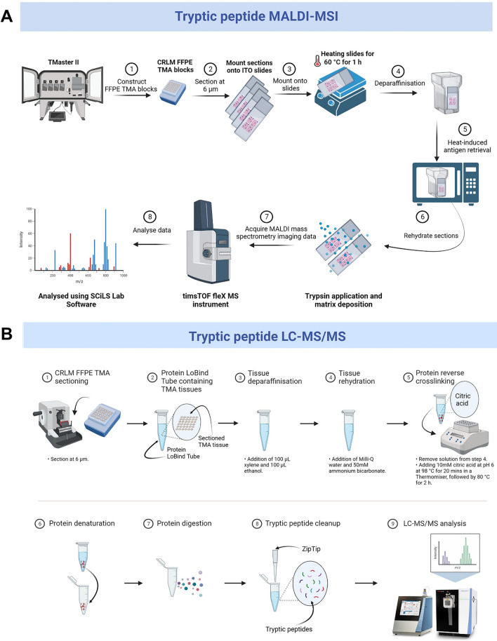
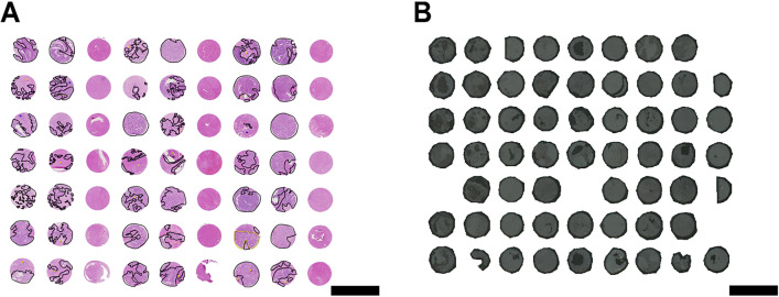
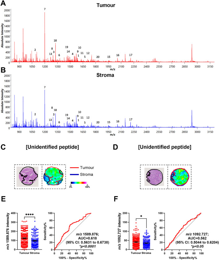
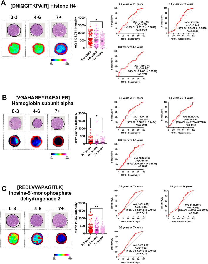
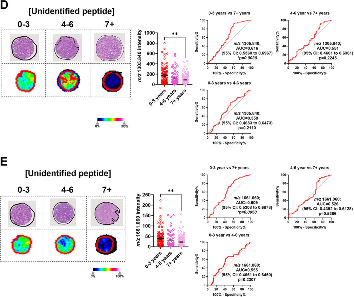
Colorectal cancer (CRC) is the second leading cause of cancer-related deaths worldwide. CRC liver metastases (CRLM) are often resistant to conventional treatments, with high rates of recurrence. Therefore, it is crucial to identify biomarkers for CRLM patients that predict cancer progression. This study utilised matrix-assisted laser desorption/ionisation mass spectrometry imaging (MALDI-MSI) in combination with liquid chromatography-tandem mass spectrometry (LC-MS/MS) to spatially map the CRLM tumour proteome. CRLM tissue microarrays (TMAs) of 84 patients were analysed using tryptic peptide MALDI-MSI to spatially monitor peptide abundances across CRLM tissues. Abundance of peptides was compared between tumour vs stroma, male vs female and across three groups of patients based on overall survival (0-3 years, 4-6 years, and 7+ years). Peptides were then characterised and matched using LC-MS/MS. A total of 471 potential peptides were identified by MALDI-MSI. Our results show that two unidentified m/z values (1589.876 and 1092.727) had significantly higher intensities in tumours compared to stroma. Ten m/z values were identified to have correlation with biological sex. Survival analysis identified three peptides (Histone H4, Haemoglobin subunit alpha, and Inosine-5'-monophosphate dehydrogenase 2) and two unidentified m/z values (1305.840 and 1661.060) that were significantly higher in patients with shorter survival (0-3 years relative to 4-6 years and 7+ years). This is the first study using MALDI-MSI, combined with LC-MS/MS, on a large cohort of CRLM patients to identify the spatial proteome in this malignancy. Further, we identify several protein candidates that may be suitable for drug targeting or for future prognostic biomarker development.

Keywords: Biomarkers; Colorectal cancer; Colorectal cancer liver metastasis; Drug targets; LC-MS/MS; MALDI-MSI.


Conflict of interest statement

The authors have no relevant financial or non-financial interests to disclose.

Figures

Fig. 1
Fig. 1
Overview of A MALDI-MSI and B LC-MS/MS workflows for spatial mapping and characterisation of tryptic peptides from CRLM FFPE tissues (n = 84). Created with BioRender.com.
Fig. 2
Fig. 2
Representative scanned images of A H&E CRLM TMA cores, and B optical CLRM TMA cores. CRLM tumour regions and CRC tumour regions (control) in A were annotated by a pathologist, as shown in black and green, respectively. Scale bars (black lines) represent 3mm.
Fig. 3
Fig. 3
Identification of tryptic peptides associated with CRLM tumour regions using MALDI-MSI and LC-MS/MS. A Sum spectra of CRLM tumour (red) and B stroma (blue) in CRLM tumour cores. C, D H&E images (left) of a representative tumour core with tumour regions annotated in black and ion intensity maps (right) of most discriminative m/z values between tumour (red) and stroma (red). Tumour cores are 1.5mm in diameter. Bar graphs showing mean ± SEM and ROC plots for E m/z 1589.876 and F m/z 1092.727. *p<0.05, ****p<0.0001. Student’s t test. Each dot represents a single tumour core.
Fig. 4
Fig. 4
Significantly abundant tryptic peptides associated with male and female CRLM tumour regions, by MALDI-MSI and LC-MS/MS. A Representative H&E and ion intensity map of tumour region annotated CRLM tumour cores for m/z 2211.113. Tumour cores are 1.5mm in diameter. AD Tryptic peptides (m/z 2211.113, 1296.682, 1570.676, and 1366.628) significantly more abundant in females relative to males. Tryptic peptides were identified as A unidentified, B Proteasome subunit beta type-9, C unidentified, and D Tryptase beta-2 Tryptase alpha/beta-1. E-H Tryptic peptides (m/z 1669.829, 1461.702, 1466.697, and 1235.618) significantly more abundant in males relative to females. These tryptic peptides were identified as E unidentified, F Carcinoembryonic antigen-related cell adhesion molecule 5, G Heat shock cognate 71 kDa protein, and H unidentified. Bar graphs showing mean ± SEM and ROC plots are presented. ****p<0.0001. Student’s t test. Each dot represents a single tumour core.
Fig. 5
Fig. 5
Significantly abundant tryptic peptides associated with male and female CRLM stroma regions, by MALDI-MSI and LC-MS/MS. A Representative H&E and ion intensity map of stroma region annotated CRLM tumour cores for m/z 1366.628. Tumour cores are 1.5 mm in diameter. AF Tryptic peptides (m/z 1366.628, 1296.682, 2211.113, 1570.676, 1661.060 and 1305.840) significantly more abundant in females relative to males. Tryptic peptides were identified as A Tryptase beta-2 Tryptase alpha/beta-1, B Proteasome subunit beta type-9, and CF unidentified. GH Tryptic peptides (m/z 1669.829 and 1461.702) significantly more abundant in males relative to females. These tryptic peptides were identified as G unidentified and H Carcinoembryonic antigen-related cell adhesion molecule 5. Bar graphs showing mean ± SEM and ROC plots are presented. ****p<0.0001. Student’s t test. Each dot represents a single tumour core.
Fig. 6
Fig. 6
Significantly abundant tryptic peptides associated with poor overall survival (0–3 years) in CRLM tumour regions. CRLM cores were subdivided based on overall survival after curative-intent surgery (0–3, 4–6, and 7+ years). Representative H&E and ion intensity maps of tumour region annotated CRLM tumour cores for m/z 1325.754, 1529.738, 1481.857, 1305.840, and 1661.060. Tumour cores are 1.5mm in diameter. These tryptic peptides were identified as A Histone H4, B Haemoglobin subunit alpha, C Inosine-5’-monophosphase dehydrogenase 2 and DE unidentified by LC-MS/MS. Bar graphs showing mean ± SEM and ROC plots are presented. *p<0.05, **p<0.01. Ordinary one-way ANOVA. Each dot represents a single tumour core.
Fig. 6
Fig. 6
Significantly abundant tryptic peptides associated with poor overall survival (0–3 years) in CRLM tumour regions. CRLM cores were subdivided based on overall survival after curative-intent surgery (0–3, 4–6, and 7+ years). Representative H&E and ion intensity maps of tumour region annotated CRLM tumour cores for m/z 1325.754, 1529.738, 1481.857, 1305.840, and 1661.060. Tumour cores are 1.5mm in diameter. These tryptic peptides were identified as A Histone H4, B Haemoglobin subunit alpha, C Inosine-5’-monophosphase dehydrogenase 2 and DE unidentified by LC-MS/MS. Bar graphs showing mean ± SEM and ROC plots are presented. *p<0.05, **p<0.01. Ordinary one-way ANOVA. Each dot represents a single tumour core.

Similar articles

Cited by

References

    1. Morgan E, et al. Global burden of colorectal cancer in 2020 and 2040: incidence and mortality estimates from GLOBOCAN. Gut. 2023;72(2):338–44. 10.1136/gutjnl-2022-327736. - PubMed
    1. Zhou H, et al. Colorectal liver metastasis: molecular mechanism and interventional therapy. Signal Transd Target Ther. 2022;7(1):70. 10.1038/s41392-022-00922-2. - PMC - PubMed
    1. Dong Y, Gruenberger T. Surgical management of colorectal liver metastases—a practical clinical approach. European Surgery. 2023;55(3):94–9. 10.1007/s10353-023-00796-w.
    1. Zhou H, et al. Colorectal liver metastasis: molecular mechanism and interventional therap. Signal Transduct Target Ther. 2022;7(1):70. 10.1038/s41392-022-00922-2. - PMC - PubMed
    1. Reinert T, et al. Circulating tumor DNA for prognosis assessment and postoperative management after curative-intent resection of colorectal liver metastases. Int J Cancer. 2022;150(9):1537–48. 10.1002/ijc.33924. - PMC - PubMed