

Diffusion-weighted imaging-based radiomics model using automatic machine learning to differentiate cerebral cystic metastases from brain abscesses
- PMID: 38492096
- PMCID: PMC10944436
- DOI: 10.1007/s00432-024-05642-4
Diffusion-weighted imaging-based radiomics model using automatic machine learning to differentiate cerebral cystic metastases from brain abscesses
Abstract
Objectives: To develop a radiomics model based on diffusion-weighted imaging (DWI) utilizing automated machine learning method to differentiate cerebral cystic metastases from brain abscesses.
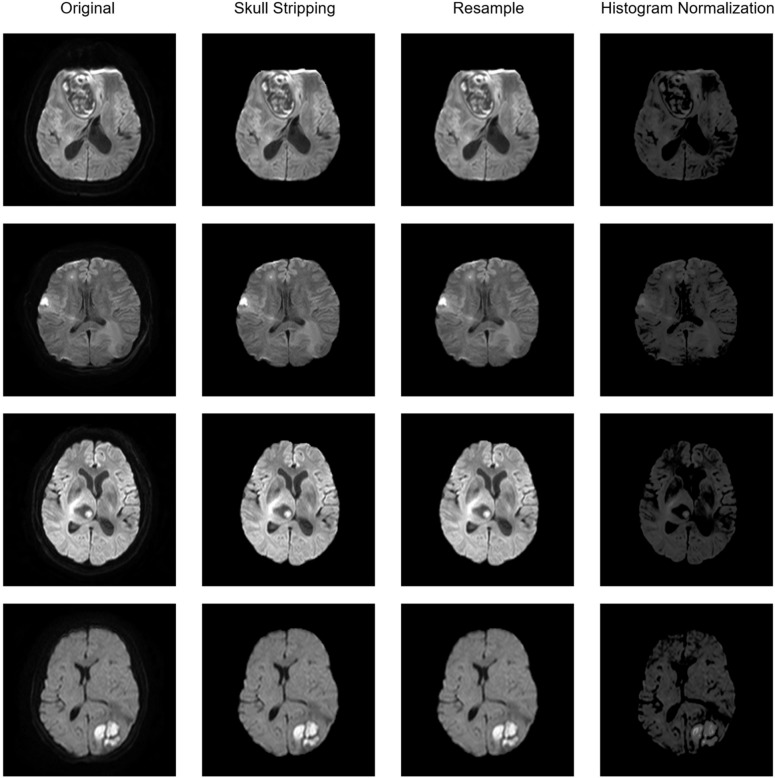
Materials and methods: A total of 186 patients with cerebral cystic metastases (n = 98) and brain abscesses (n = 88) from two clinical institutions were retrospectively included. The datasets (129 from institution A) were randomly portioned into separate 75% training and 25% internal testing sets. Radiomics features were extracted from DWI images using two subregions of the lesion (cystic core and solid wall). A thorough image preprocessing method was applied to DWI images to ensure the robustness of radiomics features before feature extraction. Then the Tree-based Pipeline Optimization Tool (TPOT) was utilized to search for the best optimized machine learning pipeline, using a fivefold cross-validation in the training set. The external test set (57 from institution B) was used to evaluate the model's performance.
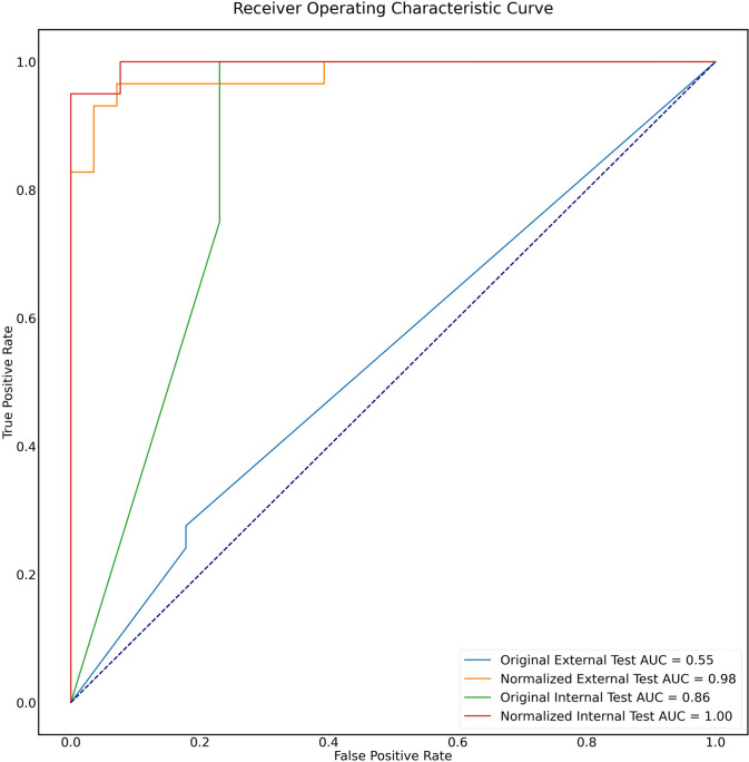
Results: Seven distinct TPOT models were optimized to distinguish between cerebral cystic metastases and abscesses either based on different features combination or using wavelet transform. The optimal model demonstrated an AUC of 1.00, an accuracy of 0.97, sensitivity of 1.00, and specificity of 0.93 in the internal test set, based on the combination of cystic core and solid wall radiomics signature using wavelet transform. In the external test set, this model reached 1.00 AUC, 0.96 accuracy, 1.00 sensitivity, and 0.93 specificity.
Conclusion: The DWI-based radiomics model established by TPOT exhibits a promising predictive capacity in distinguishing cerebral cystic metastases from abscesses.
Keywords: Brain abscesses; Cerebral cystic metastases; Image normalization; Tree-based optimization tool.
© 2024. The Author(s).
Conflict of interest statement
The authors declare no conflict of interest.
Figures


Similar articles
-
[Application of Automated Machine Learning Based on Radiomics Features of T2WI and RS-EPI DWI to Predict Preoperative T Staging of Rectal Cancer].Sichuan Da Xue Xue Bao Yi Xue Ban. 2021 Jul;52(4):698-705. doi: 10.12182/20210460201. Sichuan Da Xue Xue Bao Yi Xue Ban. 2021. PMID: 34323052 Free PMC article. Chinese.
-
Identification of testicular cancer with T2-weighted MRI-based radiomics and automatic machine learning.BMC Cancer. 2025 Mar 28;25(1):563. doi: 10.1186/s12885-025-13844-3. BMC Cancer. 2025. PMID: 40155850 Free PMC article.
-
A Comprehensive Machine Learning Benchmark Study for Radiomics-Based Survival Analysis of CT Imaging Data in Patients With Hepatic Metastases of CRC.Invest Radiol. 2023 Dec 1;58(12):874-881. doi: 10.1097/RLI.0000000000001009. Epub 2023 Jul 28. Invest Radiol. 2023. PMID: 37504498 Free PMC article.
-
Decoding Radiomics: A Step-by-Step Guide to Machine Learning Workflow in Hand-Crafted and Deep Learning Radiomics Studies.Diagnostics (Basel). 2024 Nov 5;14(22):2473. doi: 10.3390/diagnostics14222473. Diagnostics (Basel). 2024. PMID: 39594139 Free PMC article. Review.
-
The pitfalls of fixed-ratio data splitting in radiomics model performance evaluation.Abdom Radiol (NY). 2025 Apr 10. doi: 10.1007/s00261-025-04936-6. Online ahead of print. Abdom Radiol (NY). 2025. PMID: 40208285 Review.
Cited by
-
Virtual Reality for Preoperative Planning and Education in Pediatric Surgery: Preliminary Results for the Treatment of Congenital Malformations and Tumors.World J Surg. 2025 Jun;49(6):1497-1507. doi: 10.1002/wjs.12594. Epub 2025 Apr 17. World J Surg. 2025. PMID: 40246557 Free PMC article.
-
Brain Abscess Mimicking Brain Tumors: A Systematic Review of Individual Patient's Data.Asian J Neurosurg. 2025 Feb 6;20(2):291-300. doi: 10.1055/s-0045-1802623. eCollection 2025 Jun. Asian J Neurosurg. 2025. PMID: 40485794 Free PMC article.
-
Clinicodemographic and Radiological Features of Infective Ring-Enhancing Brain Lesions: A 4-Year Retrospective Study at a Tertiary Referral Center.Open Forum Infect Dis. 2025 Feb 26;12(3):ofaf095. doi: 10.1093/ofid/ofaf095. eCollection 2025 Mar. Open Forum Infect Dis. 2025. PMID: 40046884 Free PMC article.
References
-
- Alam MS, Sajjad Z, Azeemuddin M, Khan ZA, Mubarak F, Akhtar W (2012) Diffusion weighted MR imaging of ring enhancing brain lesions. J Coll Physicians Surg Pak 22(7):428–431 - PubMed
-
- Bodilsen J, Duerlund LS, Mariager T et al (2023) Clinical features and prognostic factors in adults with brain abscess. Brain 146(4):1637–1647. 10.1093/brain/awac312 - PubMed
-
- Cui Y, Yin FF (2022) Impact of image quality on radiomics applications. Phys Med Biol. 10.1088/1361-6560/ac7fd7 - PubMed
MeSH terms
LinkOut - more resources
Full Text Sources
