. 2024 Apr;22(2):207-223.
doi: 10.1007/s12021-024-09656-8. Epub 2024 Mar 16.

Updates to the Melbourne Children's Regional Infant Brain Software Package (M-CRIB-S)

Affiliations

Updates to the Melbourne Children's Regional Infant Brain Software Package (M-CRIB-S)

Chris L Adamson et al. Neuroinformatics. 2024 Apr.

Abstract

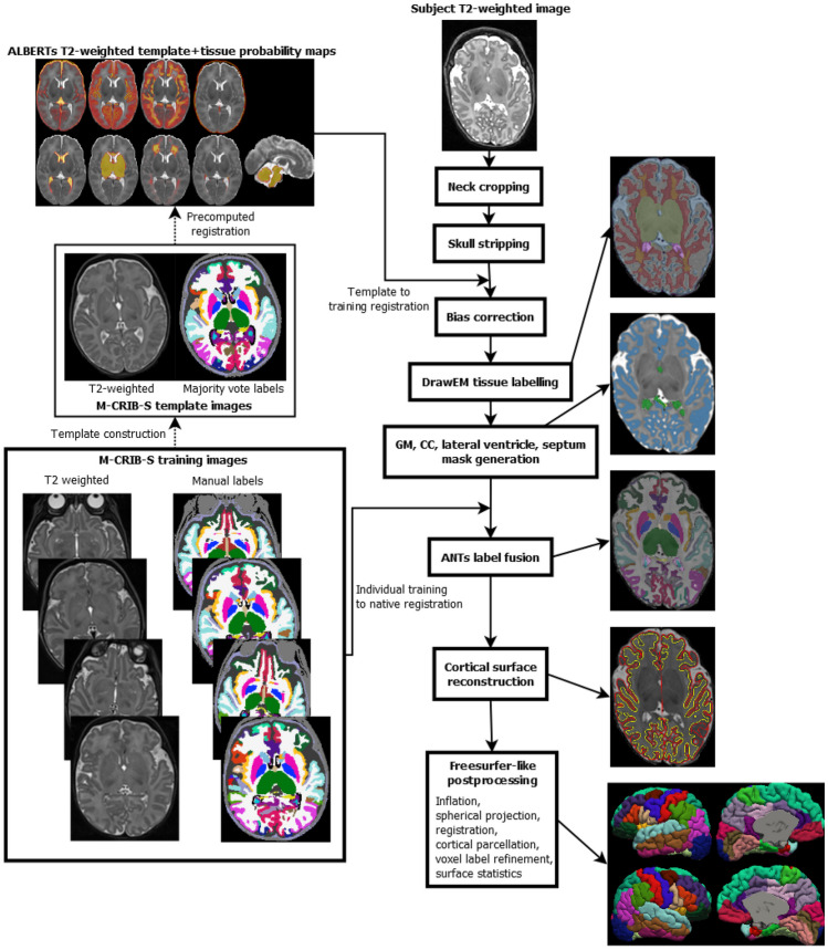
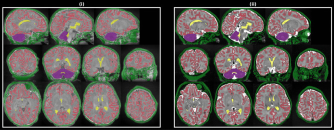
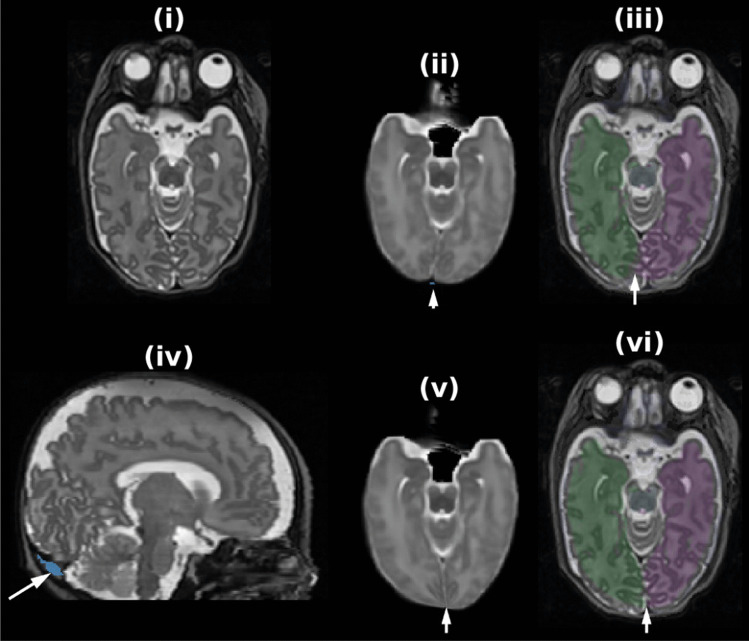
The delineation of cortical areas on magnetic resonance images (MRI) is important for understanding the complexities of the developing human brain. The previous version of the Melbourne Children's Regional Infant Brain (M-CRIB-S) (Adamson et al. Scientific Reports, 10(1), 10, 2020) is a software package that performs whole-brain segmentation, cortical surface extraction and parcellation of the neonatal brain. Available cortical parcellation schemes in the M-CRIB-S are the adult-compatible 34- and 31-region per hemisphere Desikan-Killiany (DK) and Desikan-Killiany-Tourville (DKT), respectively. We present a major update to the software package which achieves two aims: 1) to make the voxel-based segmentation outputs derived from the Freesurfer-compatible M-CRIB scheme, and 2) to improve the accuracy of whole-brain segmentation and cortical surface extraction. Cortical surface extraction has been improved with additional steps to improve penetration of the inner surface into thin gyri. The improved cortical surface extraction is shown to increase the robustness of measures such as surface area, cortical thickness, and cortical volume.

Keywords: Baby; Cortical; Gyrus; Magnetic resonance imaging; Neonate; Segmentation; Sulcus.


Conflict of interest statement

The authors declare no competing interests.

Figures

Fig. 1
Fig. 1
Depiction of some voxel-based segmentation labels for the Freesurfer-compatible M-CRIB (used in this software version) and the Freesurfer-incompatible ALBERTs (used in the previous software version) schemes that are incompatible
Fig. 2
Fig. 2
Depiction of the pipeline employed by the new version of the software. After neck cropping, skull stripping, and bias correction, the ALBERTs 40-week template and posterior fissure mask are registered to subject native space followed by DrawEM tissue labelling into 9 classes. The DrawEM tissue labels and majority vote M-CRIB labels are used to form approximate GM, CC, lateral ventricle masks. All 10 M-CRIB training intensity and label images are registered to the native image which are used to perform ANTs label fusion. Cortical surface reconstruction, using a Deformable-based method is performed. Finally, a Freesurfer-like pipeline incorporating cortical surface inflation, spherical projection, registration, parcellation with vertex- and DKT-region-wise thickness, surface area, volume measures is performed
Fig. 3
Fig. 3
One training subject’s T1-weighted (i) and T2-weighted (ii) images with structure label masks used for template construction registration colored as follows: cerebral GM (red), Cerebellum (purple), “Skull” (green), Lateral Ventricles and central CSF (yellow)
Fig. 4
Fig. 4
Example of the proposed skull stripping procedure. After BET (i) a 4-class K-means clustering is applied to the intensities within the mask (ii). The outer stripe of the scalp and other non-brain tissue is then mostly removed with a morpholgical opening, largest component, morphological closing to produce the final brain mask (iii)
Fig. 5
Fig. 5
Fissure alignment procedure whose initial input is the subject image (i) and the template image after affine registration (ii). In (iii) inspection of the left/right (purple/green) hemisphere labels reveal misalignment at the posterior fissure with the template left hemisphere penetrating the right hemisphere of the subject image (see the arrowhead). The blue regions shown in (arrowhead ii) and (arrowhead iv) show the manual/automatically delineated “posterior fissure” masks, respectively. Figures (v) and (vi) depict the result of the non-linear fissure mask + intensity warping which demonstrates the correct alignment of the template image hemispheres post-warping (arrowhead vi)
Fig. 6
Fig. 6
Depiction of approximate GM label mask creation routine. (i) T2-weighted image, (ii) DrawEM GM label overlaid (blue) with red arrows highlighting errors, (iii) sign of 2nd derivative of T2-weighted image colored as follows: negative (red, locally bright), positive (green, locally dark) or almost zero (transparent, locally uniform). (iv) Connected components of positive 2nd derivative voxels from panel (iii) colored according to the proportion of their perimeter neighboring the original GM label in panel (ii); Red and blue arrows highlight above- and below-threshold connected components, respectively. The final GM label is shown in (v), which shows the result of connected components thresholding
Fig. 7
Fig. 7
Septum pellucidum (SP) mask generation example. (i) shows the original T2-weighted subject image with the left/right SP denoted by red/blue arrowheads. (ii) shows the template left/right SP masks after registration. (iii) shows the estimated SP masks after lateral search from the masks in (ii)
Fig. 8
Fig. 8
Example of a connected component labelled by the label fusion as CSF (green and blue crosshair), relabelled as WM by being connected to the largest WM connected component
Fig. 9
Fig. 9
Label pair boundary voxel label replacement example for Superior Temporal (blue) and Supramarginal (green) label. The label of the voxels between the space where the labels meet (the red crosshair) will be replaced with the CSF label
Fig. 10
Fig. 10
WM surface construction steps. (i) With “regions.nii.gz” overlaid. The yellow circle highlights a region of interest where the proposed fix had a noticeable effect
Fig. 11
Fig. 11
Example of manual edits to fix a “pial-5” error; the error is an erroneous occlusion of the white matter (WM) surface (crosshair in (i)). In (i) the “inner” band is the final white matter surface while the outer band is the initial configuration of the pial surface after outward projection. The red edges underneath the cross hair denote vertices that are members of self-intersecting faces. The voxels are labelled as RH WM (white), LH WM (red), GM (blue), cerebellum (purple). (ii) The edited image labelfusion_dkt_edited.nii.gz with edited RH WM voxels shown as blue underneath the crosshair. (iii) Final white (yellow) and pial (red) surfaces after rerunning the surface extraction pipeline
Fig. 12
Fig. 12
Difference in variances between methods (old – new) for voxel-based structure volumes (i) and geometric properties of DKT cortical regions (ii-iv) that are as follows: surface area (ii), mean cortical thickness (iii), standard deviation of cortical thickness (iv)
Fig. 13
Fig. 13
Corpus Callosum segmentation improvements from the original software version using DrawEM (i) to the new software version using M-CRIB-S (ii) for two example subjects. The estimated CC labels are shown in purple with the ground truth, manually delineated by author C.A., shown by outline as red contours

References

    1. Adamson CL, Alexander B, Ball G, Beare R, Cheong JLY, Spittle AJ, Doyle LW, Anderson PJ, Seal ML, Thompson DK. Parcellation of the neonatal cortex using surface-based Melbourne Children’s Regional Infant Brain atlases (M-CRIB-S) Scientific Reports. 2020;10(1):10. doi: 10.1038/s41598-020-61326-2. - DOI - PMC - PubMed
    1. Alexander B, Murray AL, Loh WY, Matthews LG, Adamson C, Beare R, Chen J, Kelly CE, Rees S, Warfield SK, Anderson PJ, Doyle LW, Spittle AJ, Cheong JL, Seal ML, Thompson DK. A new neonatal cortical and subcortical brain atlas: The Melbourne Children's Regional Infant Brain (M-CRIB) atlas. NeuroImage. 2017;147:841–851. doi: 10.1016/j.neuroimage.2016.09.068. - DOI - PubMed
    1. Alexander, B., Loh, W. Y., Matthews, L. G., Murray, A. L., Adamson, C., Beare, R., Chen, J., Kelly, C. E., Anderson, P. J., Doyle, L. W., Spittle, A. J., Cheong, J. L. Y., Seal, M. L., & Thompson, D. K. (2019). Desikan-Killiany-Tourville atlas compatible version of M-CRIB neonatal parcellated whole brain atlas: The M-CRIB 2.0. Frontiers in Neuroscience, 13, 34. 10.3389/fnins.2019.00034 - PMC - PubMed
    1. Avants BB, Epstein CL, Grossman M, Gee JC. Symmetric diffeomorphic image registration with cross-correlation: Evaluating automated labeling of elderly and neurodegenerative brain. Medical Image Analysis. 2008;12(1):26–41. doi: 10.1016/j.media.2007.06.004. - DOI - PMC - PubMed
    1. Beare, R. J., Chen, J., Kelly, C. E., Alexopoulos, D., Smyser, C. D., Rogers, C. E., Loh, W. Y., Matthews, L. G., Cheong, J. L. Y., Spittle, A. J., Anderson, P. J., Doyle, L. W., Inder, T. E., Seal, M. L., & Thompson, D. K. (2016). Neonatal brain tissue classification with morphological adaptation and unified segmentation. Frontiers in Neuroinformatics, 10, 12. https://www.frontiersin.org/article/10.3389/fninf.2016.00012 - DOI - PMC - PubMed