. 2024 Mar 16;30(1):39.
doi: 10.1186/s10020-024-00784-0.

Involvement of Fgf2-mediated tau protein phosphorylation in cognitive deficits induced by sevoflurane in aged rats

Affiliations

Involvement of Fgf2-mediated tau protein phosphorylation in cognitive deficits induced by sevoflurane in aged rats

Xin Xie et al. Mol Med. .

Abstract

Objective: Anesthetics have been linked to cognitive alterations, particularly in the elderly. The current research delineates how Fibroblast Growth Factor 2 (Fgf2) modulates tau protein phosphorylation, contributing to cognitive impairments in aged rats upon sevoflurane administration.

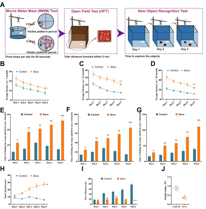
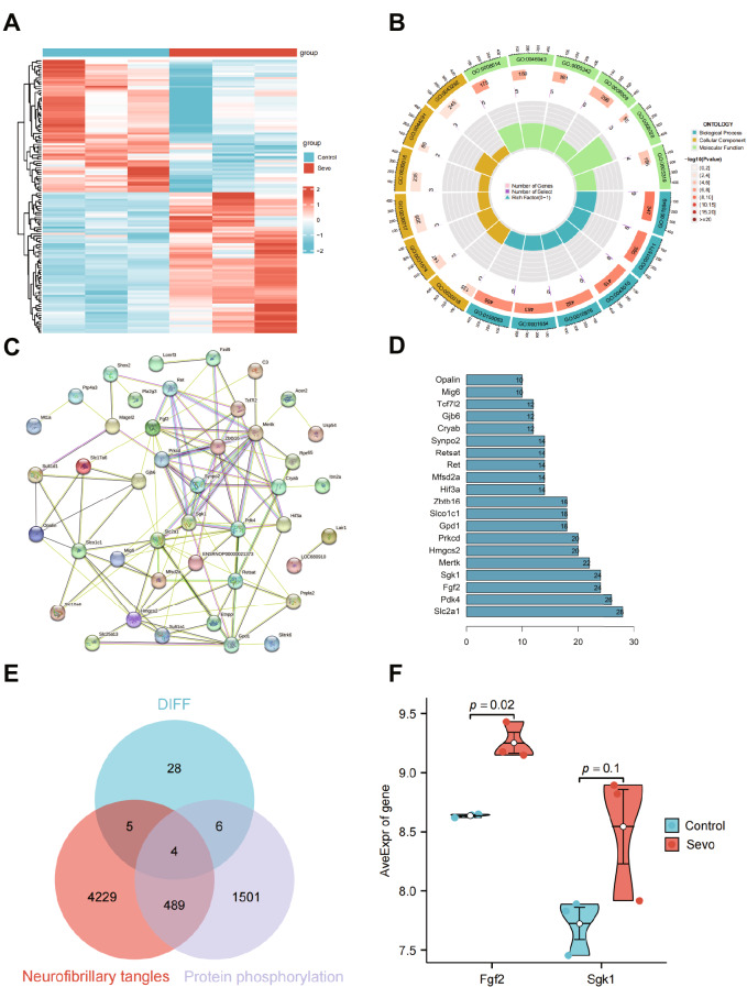
Methods: Rats aged 3, 12, and 18 months were subjected to a 2.5% sevoflurane exposure to form a neurotoxicity model. Cognitive performance was gauged, and the GEO database was employed to identify differentially expressed genes (DEGs) in the 18-month-old cohort post sevoflurane exposure. Bioinformatics tools, inclusive of STRING and GeneCards, facilitated detailed analysis. Experimental validations, both in vivo and in vitro, examined Fgf2's effect on tau phosphorylation.

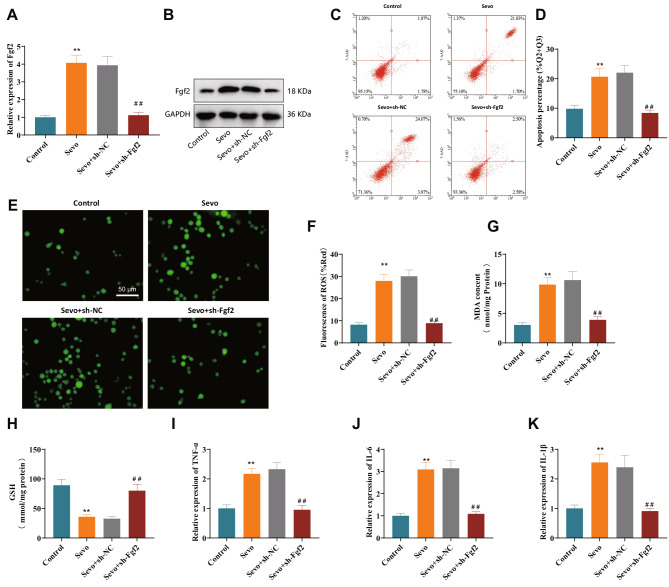
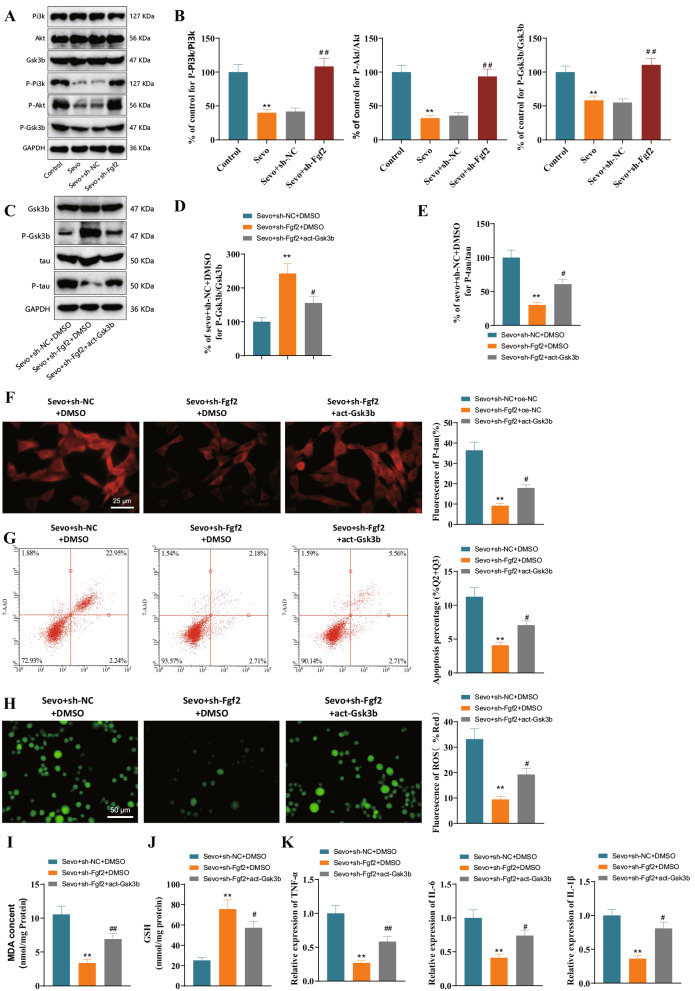
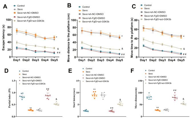
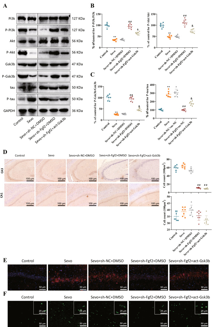
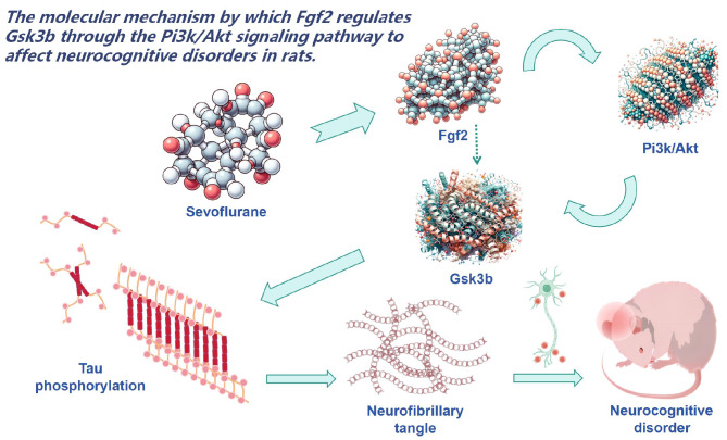
Results: Sevoflurane notably altered cognitive behavior in older rats. Out of 128 DEGs discerned, Fgf2 stood out as instrumental in regulating tau protein phosphorylation. Sevoflurane exposure spiked Fgf2 expression in cortical neurons, intensifying tau phosphorylation via the PI3K/AKT/Gsk3b trajectory. Diminishing Fgf2 expression correspondingly curtailed tau phosphorylation, neurofibrillary tangles, and enhanced cognitive capacities in aged rats.

Conclusion: Sevoflurane elicits a surge in Fgf2 expression in aging rats, directing tau protein phosphorylation through the PI3K/AKT/Gsk3b route, instigating cognitive aberrations.

Keywords: Aged rats; Cognitive impairment; Fgf2; Gsk3b; Sevoflurane; Tau protein phosphorylation; Transcriptomic sequencing.


Conflict of interest statement

The authors declare that they have no competing interests.

Figures

Fig. 1
Fig. 1
Effects of sevoflurane exposure on rats’ learning, cognition, and motor ability. Note: (A) Behavioral Experiment Design and Timeline; (BD) Line graphs showing the changes in escape latency during different days in the water maze experiment for rats in different treatment groups at 3 months (Fig. B), 12 months (Fig. C), and 18 months (Fig. D); (DF) Bar graphs showing the time required for rats in different treatment groups at 3 months (Fig. E), 12 months (Fig. F), and 18 months (Fig. G) to find the target platform on different days in the water maze experiment; (H) Open field test measuring the time for 18-month-aged rats in different treatment groups to initiate movement; (I) Open field test measuring the total distance traveled by 18-month-aged rats in different treatment groups; (J) Index of novel object recognition in the open field test. P < 0.05, P < 0.01, **P < 0.001, all significances are compared between the Sevo and Control groups; each group consisted of 7 rats
Fig. 2
Fig. 2
Core factors involved in sevoflurane-induced cognitive impairment identified by bioinformatics analysis. Note: (A) Heatmap showing the differentially expressed genes (DEGs) between standard control samples (Control group, n = 3) and sevoflurane-induced neurotoxicity rat samples (Sevo group, n = 3) in GSE141242 (LogFC > 0.5, P value < 0.05). (B) Functional enrichment analysis results of the DEGs at the biological process (BP), cellular component (CC), and molecular function (MF) levels. (C) Protein-protein interaction (PPI) network was constructed for the 128 encoded proteins of the DEGs identified in GSE141242. (D) Top 20 genes ranked by the number of connections in the PPI network. (E) The intersection between 43 DEGs (DIFF) identified in GSE141242 and genes associated with neurofibrillary tangles and protein phosphorylation. (F) Analysis of the differential expression of critical genes Fgf2 and Sgk1 using the Welch to test statistical method
Fig. 3
Fig. 3
Effects of silencing Fgf2 on neuronal apoptosis, oxidative stress injury, and neuroinflammation in the in vitro sevoflurane model. Note: (A) RT-qPCR analysis of Fgf2 mRNA expression in sevoflurane model neurons; (B) Western blot analysis of Fgf2 protein expression in sevoflurane model neurons; (C) Flow cytometry analysis of apoptosis in primary cortical neurons from different groups (Q2 and Q3 quadrants); (D) Statistical analysis of the percentage of apoptosis in different groups of primary cortical neurons (Q2 and Q3 quadrants); (EF) Generation of reactive oxygen species (ROS) in different groups of primary cortical neurons (green fluorescence represents ROS activity marker, scale bar = 50 μm); (G) Measurement of malondialdehyde (MDA) content in different groups of primary cortical neurons using the TBARS assay; (H) Measurement of glutathione (GSH) content in different groups of primary cortical neurons using the glutathione assay kit; (IK) RT-qPCR analysis of TNF-α, IL-6, and IL-1β expression in different groups of primary cortical neurons. ** represents a difference compared to the Control group or Sevo + sh-NC group (P < 0.01), ## represents a difference compared to the Sevo + sh-Fgf2 group (P < 0.01), and the cell experiments were repeated 3 times
Fig. 4
Fig. 4
Fgf2 regulates PI3K/AKT/Gsk3b pathway to promote tau protein phosphorylation in Sevo model neurons. Note: (A) Western blot analysis of protein expression of PI3K, AKT, Gsk3b, P-PI3K, P-AKT, and P-Gsk3b in Sevo model neurons after silencing Fgf2. (B) Statistical analysis of protein expression ratios of P-PI3K/PI3K, P-AKT/AKT, P-Gsk3b/Gsk3b in Sevo model neurons after silencing Fgf2. (C) Western blot analysis of protein expression of Gsk3b, P-Gsk3b, tau, and P-tau in Sevo model neurons after silencing Fgf2 and Gsk3b activation. (D) Statistical analysis of protein expression ratio of P-Gsk3b/Gsk3b. (E) Statistical analysis of protein expression ratio of P-tau/tau. (F) Immunofluorescence detection of P-tau content in primary cortical neurons of various groups after silencing Fgf2 and Gsk3b activation. (G) Flow cytometry analysis of apoptosis and proportion (Q2 and Q3 quadrants) in primary cortical neurons of various groups after silencing Fgf2 and Gsk3b activation. (H) Measurement of ROS production in primary cortical neurons of various groups after silencing Fgf2 and Gsk3b activation. (I) TBARS assay to detect MDA levels in primary cortical neurons of various groups after silencing Fgf2 and Gsk3b activation. (J) Glutathione assay to detect GSH levels in primary cortical neurons of various groups after silencing Fgf2 and Gsk3b activation. (K) RT-qPCR analysis of TNF-α, IL-6, and IL-1β expression in primary cortical neurons of various groups after silencing Fgf2 and Gsk3b activation. ** indicates comparison with Control group or Sevo + sh-NC + DMSO group, P < 0.01; # indicates comparison with Sevo + sh-Fgf2 + DMSO group, P < 0.05; ## indicates comparison with Sevo + sh-NC group, P < 0.01. Cell experiments were repeated three times
Fig. 5
Fig. 5
Fgf2 affects motor and learning impairments in aged rats. Note: (A) Line graph showing the escape latency duration of rats in different treatment groups in the water maze experiment on different days. (B) Distance traveled by rats in different treatment groups to reach the specific platform on different days in the water maze experiment. (C) Time taken by rats in different treatment groups to reach the specific platform on different days in the water maze experiment. (D) Exploration index of rats for the novel object in the open field test. (E) Starting time of rat movement in the open field test for different treatment groups. (F) Total distance traveled by rats in the open field test for different treatment groups. * indicates comparison with Control group, P < 0.05; ** indicates comparison with Control group, P < 0.01; ## indicates comparison with Sevo + sh-NC + DMSO group, P < 0.01; & indicates comparison with Sevo + sh-Fgf2 + DMSO group, P < 0.05. Each group consisted of seven rats
Fig. 6
Fig. 6
Fgf2 promotes abnormal accumulation of tau protein and neurofibrillary tangle formation by regulating Gsk3b. Note: (A) Western blot analysis of PI3K, P-PI3K, AKT, P-AKT, Gsk3b, P-Gsk3b, tau, and P-tau protein expression in Sevo model rats. (B) Statistical analysis of P-PI3K/PI3K and P-AKT/AKT protein expression ratios. (C) Statistical analysis of P-Gsk3b/Gsk3b and P-tau/tau protein expression ratios. (D) Immunohistochemistry and corresponding quantitative analysis of Fgf2 in the CA3 and CA1 regions confirmed the successful knockdown of Fgf2 in the CA3 region. (E) Immunofluorescence detection of P-tau content in hippocampal tissues of Sevo model rats. (F) Immunofluorescence detection of neurofibrillary tangles in hippocampal tissues of Sevo model rats. Sevo group compared to Control group, **P < 0.01; ## indicates comparison between Sevo + sh-Fgf2 + DMSO or Sevo + sh-Fgf2 + act-Gsk3b group to the Sevo + sh-NC + DMSO groups, P < 0.01; & indicates comparison between Sevo + sh-Fgf2 + act-Gsk3b and Sevo + sh-Fgf2 + DMSO groups, P < 0.05. Each treatment group consisted of seven rats, and all experiments were repeated three times
Fig. 7
Fig. 7
Molecular mechanisms underlying the effect of Fgf2 on Gsk3b and its impact on cognitive impairment in rats

Similar articles

Cited by

References

    1. Amirghasemi F, Adjei-Sowah E, Pockaj BA, Nikkhah M. Microengineered 3D Tumor models for Anti-cancer Drug Discovery in female-related cancers. Ann Biomed Eng. 2021;49(8):1943–72. doi: 10.1007/s10439-020-02704-9. - DOI - PubMed
    1. Andreollo NA, Santos EF, Araújo MR, Lopes LR. Rat’s age versus human’s age: what is the relationship? Arq Bras Cir Dig. 2012;25(1):49–51. doi: 10.1590/s0102-67202012000100011. - DOI - PubMed
    1. Cai H, Luo Y, Yan X et al. The Mechanisms of Bushen-Yizhi Formula as a Therapeutic Agent against Alzheimer’s Disease. Sci Rep. 2018;8(1):3104. Published 2018 Feb 15. 10.1038/s41598-018-21468-w. - PMC - PubMed
    1. Cai ZP, Cao C, Guo Z, et al. Coeloglossum viride var. Bracteatum extract attenuates staurosporine induced neurotoxicity by restoring the FGF2-PI3K/Akt signaling axis and Dnmt3. Heliyon. 2021;7(7):e07503. doi: 10.1016/j.heliyon.2021.e07503. - DOI - PMC - PubMed
    1. Cai H, Pang Y, Wang Q et al. Proteomic profiling of circulating plasma exosomes reveals novel biomarkers of Alzheimer’s disease. Alzheimers Res Ther. 2022;14(1):181. Published 2022 Dec 5. 10.1186/s13195-022-01133-1. - PMC - PubMed

MeSH terms