. 2024 Mar 16;15(3):223.
doi: 10.1038/s41419-024-06591-z.

Casein kinase 2 phosphorylates and induces the SALL2 tumor suppressor degradation in colon cancer cells

Affiliations

Casein kinase 2 phosphorylates and induces the SALL2 tumor suppressor degradation in colon cancer cells

V E Hermosilla et al. Cell Death Dis. .

Abstract

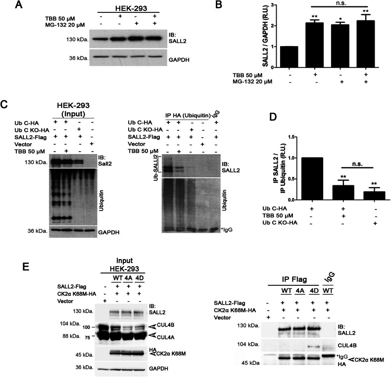
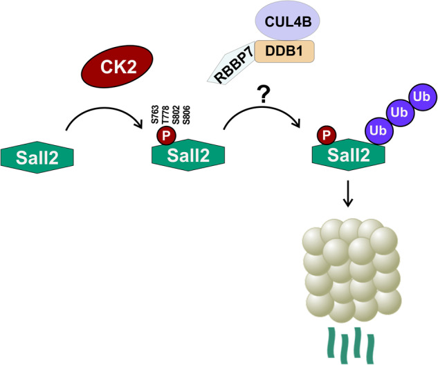
Spalt-like proteins are Zinc finger transcription factors from Caenorhabditis elegans to vertebrates, with critical roles in development. In vertebrates, four paralogues have been identified (SALL1-4), and SALL2 is the family's most dissimilar member. SALL2 is required during brain and eye development. It is downregulated in cancer and acts as a tumor suppressor, promoting cell cycle arrest and cell death. Despite its critical functions, information about SALL2 regulation is scarce. Public data indicate that SALL2 is ubiquitinated and phosphorylated in several residues along the protein, but the mechanisms, biological consequences, and enzymes responsible for these modifications remain unknown. Bioinformatic analyses identified several putative phosphorylation sites for Casein Kinase II (CK2) located within a highly conserved C-terminal PEST degradation motif of SALL2. CK2 is a serine/threonine kinase that promotes cell proliferation and survival and is often hyperactivated in cancer. We demonstrated that CK2 phosphorylates SALL2 residues S763, T778, S802, and S806 and promotes SALL2 degradation by the proteasome. Accordingly, pharmacological inhibition of CK2 with Silmitasertib (CX-4945) restored endogenous SALL2 protein levels in SALL2-deficient breast MDA-MB-231, lung H1299, and colon SW480 cancer cells. Silmitasertib induced a methuosis-like phenotype and cell death in SW480 cells. However, the phenotype was significantly attenuated in CRISPr/Cas9-mediated SALL2 knockout SW480 cells. Similarly, Sall2-deficient tumor organoids were more resistant to Silmitasertib-induced cell death, confirming that SALL2 sensitizes cancer cells to CK2 inhibition. We identified a novel CK2-dependent mechanism for SALL2 regulation and provided new insights into the interplay between these two proteins and their role in cell survival and proliferation.


Conflict of interest statement

The authors declare no competing interests.

Figures

Fig. 1
Fig. 1. SALL2 protein is a CK2 substrate in vitro.
A Scheme of human SALL2 protein. Zinc Finger motifs are highlighted in dark gray, and a putative C-terminal PEST motif is shown in green next to the DNA-binding domain. Putative phosphorylation sites matching the CK2 consensus sequence are indicated in red. Alignment of protein sequences from Homo sapiens (NP_005398.2), Mus musculus (NP_056587.2), and Rattus Norvegicus (NP_001100732.1) with Clustal omega [92] indicated that this region is highly conserved. B Endogenous SALL2 and CK2α interact in HEK 293T cells. Immunoprecipitations (IP) were performed using anti-SALL2 antibody (Bethyl Lab.), SN supernatant. C SALL2-Flag protein was co-expressed with HA-tagged wild-type CK2α or the K68M dominant-negative mutant. Immunoprecipitation assays were performed using Flag or HA antibodies. s.e., short exposure; l.e., long exposure. D SALL2-Flag was overexpressed in Flp-In™ T-REx™ U2OS cells, followed by immunoprecipitation for in vitro phosphorylation reactions. Different forms of recombinant CK2 were used, including the CK2αβ and CK2α’β holo forms and the CK2α and CK2α’ monomeric subunits, all of which phosphorylate SALL2 in vitro. Ctrl. Resin, immunoprecipitation without antibody; λ-PPase, dephosphorylation with λ-phosphatase prior to the in vitro kinase reaction. E Quantification of (D). R.U. relative units to the basal condition; error bars equal SEM; *p < 0.05, **p < 0.01, ***p < 0.001, n.s. non-significant p-value (one-way ANOVA plus Tukey’s multiple comparison test).
Fig. 2
Fig. 2. SALL2 C-terminal PEST motif is phosphorylated in a CK2-dependent manner.
A Scheme of ΔN SALL2-Flag protein lacking most of the N-terminal region, but retaining a putative N-terminal localization signal (NLS). Zinc Finger motifs are highlighted in dark gray and a putative C-terminal PEST motif is shown in green. P, Putative CK2 phosphorylation sites; FL full legth. B Ectopic expression of full-length and ΔN SALL2-Flag in Flp-In™ T-REx™ U2OS cells. C Assessment of CK2 inhibition after 6 h of treatment with Silmitasertib (CX-4945) at a final concentration of 30 µM in Flp-In™ T-REx™ U2OS cells. Phosphorylation of the CK2 substrates X-Ray Repair Cross Complementing 1 (XRCC1), Eukaryotic Translation Initiation Factor 2 beta (eIF2β), and CK2β in the indicated residues was evaluated [–98]. Phosphorylation of eIF2β (p- eIF2β) was used as a positive control of CK2 inhibition. *, non-specific band. D Phosphorylated peptides identified by Mass Spectrometry after sequential in-gel digestion. In vitro basal, in vitro kinase reaction without addition of recombinant enzyme; in vitro CK2α, in vitro kinase reaction with recombinant CK2α; cell culture (U2OS + DMSO) and cell culture (U2OS+ Silmitarsertib) represent the conditions where SALL2 was immunoprecipitated from DMSO or Silmitarsertib-treated Flp-In™ T-REx™ U2OS cells, respectively. Representative spectra of fragments carrying phosphorylation of S806 after basal (E) and CK2α-added (F) in vitro kinase reactions are shown. Representative spectra of S763 (G) and T778 (H)-containing fragments under DMSO treatment show that both residues are phosphorylated under control conditions in Flp-In™ T-REx™ U2OS cells.
Fig. 3
Fig. 3. CK2 activity downregulates SALL2 stability.
A HEK 293 cells were treated with 50 µM of the CK2 inhibitor, TBB. Cells were fixed after 7 h and subjected to immunofluorescence. Endogenous SALL2 is shown in green, whilst endogenous CK2 is stained in red. Both proteins share a nuclear localization. Negative control corresponds to no incubation with primary antibody. B Representative graph for the quantification of (A), indicating that pharmacological inhibition of CK2 resulted in an augmented immunodetection of SALL2. A.U. arbitrary units; n > 100 cells per replicate; error bars equal SEM; ***p < 0.001 (T-test). C Representative western blot of HEK 293 cells treated for 3 h with various concentrations of the specific CK2 inhibitor, Silmitasertib. D SALL2-Flag protein was co-expressed with HA-tagged wild-type CK2α or the K68M dominant-negative mutant in HEK 293 cells. Stability assays were performed using 50 µg/ml of the protein synthesis inhibitor, cycloheximide (CHX), for the indicated time-points. E Quantification of SALL2 stability when co-expressed with wild-type CK2α versus the dominant-negative kinase (K68M), normalized against GAPDH. R.U., relative units; error bars represent SEM; **p < 0.01, ***p < 0.001, n = 3 (two-way ANOVA plus Bonferroni correction).
Fig. 4
Fig. 4. CK2 activity promotes ubiquitylation and proteasome-mediated degradation of SALL2.
A HEK 293 cells were pretreated with either DMSO (vehicle) or TBB (50 µM) for 3 h, followed by incubation with DMSO (vehicle) or the proteasome inhibitor, MG-132 (20 µM), with or without TBB, for 6 additional hours. Endogenous SALL2 protein levels were quantified and normalized against GAPDH as shown in (B). R.U., relative units; error bars equal SEM; *p < 0.05, **p < 0.01, n.s. non-significant p-value (one-way ANOVA plus Tukey’s multiple comparison test). C, D Pharmacological inhibition of CK2 impairs ubiquitylation of SALL2 under proteasome inhibition. SALL2-Flag and Ubiquitin C-HA were co-expressed, and HEK 293 cells were treated as described in (A). HA-tagged ubiquitin was immunoprecipitated to enrich the pool of ubiquitylated proteins. SALL2 protein was immunodetected and quantified against Ubiquitin signal. UbC KO-HA, Ubiquitin C with mutated lysine residues; R.U. relative units; error bars represent SEM; **p < 0.01, n.s. non-significant p-value (one-way ANOVA plus Tukey’s multiple comparison test). E CK2α K68M was co-expressed with either wild-type (WT), non-phosphorylatable (4A), or phosphomimetic (4D) forms of Flag-tagged SALL2 in HEK 293 cells. SALL2 was immunoprecipitated, and interaction with endogenous CUL4B was evaluated. Endogenous CUL4B interacts with phosphomimetic SALL2 protein (4D) but not with the non-phosphorylatable (4A) or wild-type (WT) forms of SALL2.
Fig. 5
Fig. 5. Pharmacological inhibition of CK2 restores SALL2 protein levels in cancer cell lines.
A, B Silmitarsertib increases SALL2 protein levels in MDA-MB-231 triple-negative breast cancer cells. MDA-MB-231 cells were treated with increasing concentrations of Silmitarsertib for two or 6 h. SALL2 protein levels were assessed by western blot (A), quantified, and normalized against β-actin (B). R.U., relative units; error bars equal SEM; *p < 0.05, (one-way ANOVA plus Tukey’s multiple comparison test). C qRT-PCR of SALL2 normalized to 18S rRNA indicated that augmented transcription is not responsible for the upregulation of SALL2 protein after 2 h of treatment with Silmitasertib at the indicated concentrations. MDA-MB-231 cells (D) and SW480 colon cancer cells (F) were subjected to CK2 inhibition with Silmitarsertib (25 μM) and collected at the indicated time points. In both cases, SALL2 exhibited a transient upregulation when normalized to β-Actin (E, G). In addition, cleavage of PARP occurred after long-term treatment with Silmitasertib. R.U., relative units; error bars equal SEM; *p < 0.05, **p < 0.01 (repeated measures ANOVA plus Tukey’s multiple comparison test).
Fig. 6
Fig. 6. Cytotoxic effects of Silmitarsertib are decreased in SALL2-deficient SW480 cells and Sall2-deficient tumor-derived organoids.
A IC50 curve for SW480 SALL2 wild-type (clone G10), and SALL2 knockout (clone G8) cells treated with Silmitarsertib (0–40 μM) for 24 h. The percentage of cell survival is plotted against the logarithm of treatment concentrations. Data points are the means ± SD of duplicate determinations of triplicate measurements. B Real-time cytotoxic assay. The SW480 clones were treated with 25 μM Silmitarsertib and photographed every 1 h for 24 h, using INCUCYTE S3 Live-Cell Analysis System with 20× objective (A representative figure is shown). The dead cells were counted automatically every 1 h for 24 h, and the Cell death percentage was quantified with a cell-by-cell module and normalized by confluence using IncuCyte v2019B software. C Percentage of SW480 cells with formation of vacuoles after Silmitarsertib treatment. D Analysis of the number of vacuoles per cell. Each point in the graph represents an independent cell. E Size of the vacuoles observed after incubation with Silmitarsertib. Each point in the graph represents independent vacuoles observed in the indicated conditions. Analyses of Silmitarsertib-induced vacuoles in C, D, E were performed using ImageJ and Prism GraphPad software, and no less than 100 cells were counted and measured per condition. F IC50 curve for AKP control and AKP SALL2 knockout tumor-derived organoids treated with Silmitarsertib (0-100 μM) for 24 h. The percentage of cell survival is plotted against the logarithm of treatment concentrations. Data points are the means ± SD of duplicate determinations of triplicate measurements.
Fig. 7
Fig. 7. Proposed model for CK2-dependent regulation of SALL2.
CK2 activity promotes the phosphorylation of SALL2 in S763, T802, S802 and S806. Phosphorylated SALL2 interacts with CUL4B, a component of the Cullin-Ring Ubiquitin-Ligase Complexes (CRLs), being subjected to ubiquitylation followed by proteasomal degradation. Pharmacological inhibition of CK2 impairs this process, triggering the accumulation of SALL2 in cancer cells.

References

    1. de Celis JF, Barrio R. Regulation and function of Spalt proteins during animal development. Int J Dev Biol. 2009;53:1385–98. doi: 10.1387/ijdb.072408jd. - DOI - PubMed
    1. Kohlhase J. SALL1 mutations in Townes-Brocks syndrome and related disorders. Hum Mutat. 2000;16:460–6. doi: 10.1002/1098-1004(200012)16:6<460::AID-HUMU2>3.0.CO;2-4. - DOI - PubMed
    1. Kohlhase J, Hausmann S, Stojmenovic G, Dixkens C, Bink K, Schulz-Schaeffer W, et al. SALL3, a new member of the human spalt-like gene family, maps to 18q23. Genomics. 1999;62:216–22. doi: 10.1006/geno.1999.6005. - DOI - PubMed
    1. Borozdin W, Boehm D, Leipoldt M, Wilhelm C, Reardon W, Clayton-Smith J, et al. SALL4 deletions are a common cause of Okihiro and acro-renal-ocular syndromes and confirm haploinsufficiency as the pathogenic mechanism. J Med Genet. 2004;41:7–14. doi: 10.1136/jmg.2004.019901. - DOI - PMC - PubMed
    1. Paradisi I, Arias S. IVIC syndrome is caused by a c.2607delA mutation in the SALL4 locus. Am J Med Genet Part A. 2007;143:326–32. doi: 10.1002/ajmg.a.31603. - DOI - PubMed

Publication types

LinkOut - more resources