. 2024 Mar 16;7(1):336.
doi: 10.1038/s42003-024-06023-w.

Sequence-structure-function characterization of the emerging tetracycline destructase family of antibiotic resistance enzymes

Affiliations

Sequence-structure-function characterization of the emerging tetracycline destructase family of antibiotic resistance enzymes

Kevin S Blake et al. Commun Biol. .

Abstract

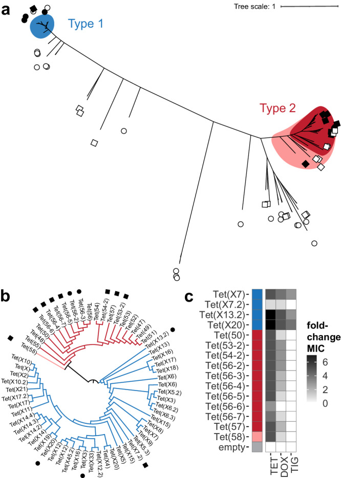
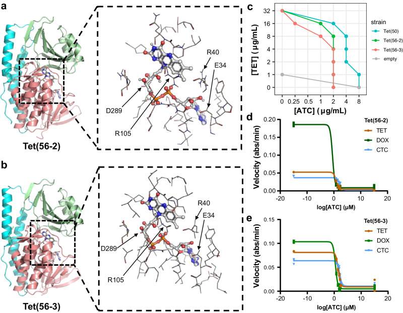
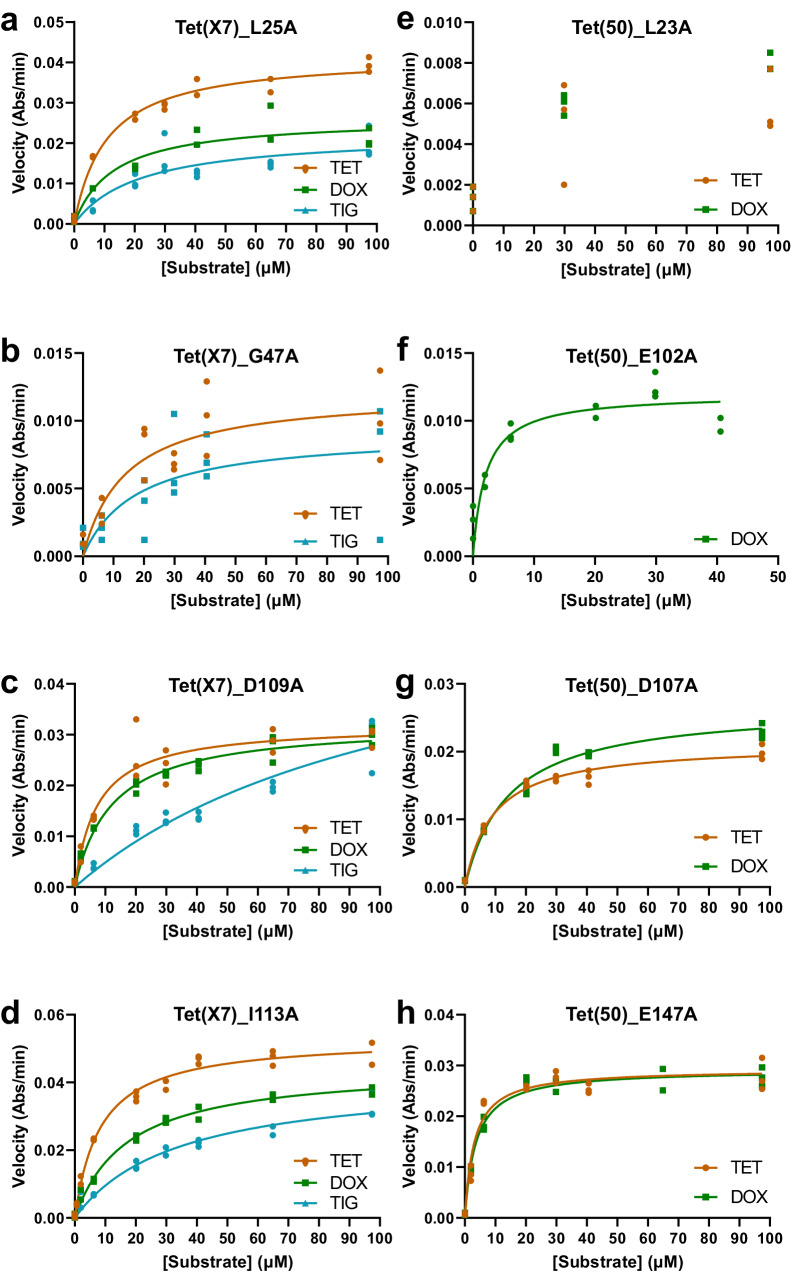
Tetracycline destructases (TDases) are flavin monooxygenases which can confer resistance to all generations of tetracycline antibiotics. The recent increase in the number and diversity of reported TDase sequences enables a deep investigation of the TDase sequence-structure-function landscape. Here, we evaluate the sequence determinants of TDase function through two complementary approaches: (1) constructing profile hidden Markov models to predict new TDases, and (2) using multiple sequence alignments to identify conserved positions important to protein function. Using the HMM-based approach we screened 50 high-scoring candidate sequences in Escherichia coli, leading to the discovery of 13 new TDases. The X-ray crystal structures of two new enzymes from Legionella species were determined, and the ability of anhydrotetracycline to inhibit their tetracycline-inactivating activity was confirmed. Using the MSA-based approach we identified 31 amino acid positions 100% conserved across all known TDase sequences. The roles of these positions were analyzed by alanine-scanning mutagenesis in two TDases, to study the impact on cell and in vitro activity, structure, and stability. These results expand the diversity of TDase sequences and provide valuable insights into the roles of important residues in TDases, and flavin monooxygenases more broadly.


Conflict of interest statement

The authors declare no competing interests.

Figures

Fig. 1
Fig. 1. Characterization of novel TDases identified using profile HMMs.
a Unrooted maximum-likelihood tree of tested HMM-predicted sequences and characterized TDase sequences. Generated with RAxML. Colored regions represent the type 1 clade (blue), the type 2 clade before HMM screening (red), and the expanded type 2 clade after HMM screening (pink). Circles indicate hmm01-predicted sequences: filled circle = functional (i.e. confers resistance to tetracycline); empty circle = does not confer resistance. Squares indicate hmm02-predicted sequences: filled square = functional; empty square = does not confer resistance. No circle or square = previously characterized TDase. b Maximum-likelihood cladogram of non-redundant characterized TDase sequences with branch lengths ignored. Generated with RAxML. Nodes annotated with name assigned in original report. Branch color indicates TDase clade (type 1 = blue; type 2 = red; type 2 after HMM screening = pink). Filled circles indicate functional sequences from hmm01, filled squares from hmm02. c Heatmap of MICs of newly identified TDase genes. Antimicrobial susceptibility testing was performed by microbroth dilution on sequences predicted by profile HMMs. Each box represents the consensus MIC value determined for three technical replicates, transformed as foldchange over the MIC values for the empty vector control. Colored box next to the protein names indicates the TDase type it belongs to (type 1 = blue; type 2 = red; type 2 after HMM screening = pink). Empty vector control, Tet(X7), and Tet(50) MICs included for reference. Acronyms = tetracycline (TET), doxycycline (DOX), tigecycline (TIG).
Fig. 2
Fig. 2. Steady-state kinetic plots of tetracycline inactivation by HMM-identified enzymes.
Steady-state kinetic plots of velocity vs substrate concentrations for the TDase-catalyzed degradation of tetracycline antibiotics were fit to the Michaelis-Menten equation. The previously characterized TDases (a) Tet(50) and (b) Tet(X7) were recharacterized here as reference to the newly identified type 2 TDases (c) Tet(56-2), (d) Tet(56-3), (e) Tet(56-4), (f) Tet(56-5), (g) Tet(56-6), (h) Tet(56-7), and (i) Tet(58), as well as the new type 1 TDase (j) Tet(X13.2). Acronyms = tetracycline (TET), doxycycline (DOX), and tigecycline (TIG). Error values represent the standard deviations for three independent trials.
Fig. 3
Fig. 3. X-ray structures and inhibition of type 2 TDases from Legionella spp.
X-ray structures of (a) Tet(56-2) (PDB 8TWG) and (b) Tet(56-3) (PDB 8TWF). The FAD-binding domain (FBD) is colored salmon, the substrate-binding domain (SBD) is colored pale green, and the C-terminal helix is colored cyan. Residues within 5 Å sphere around the bound co-factor, FAD, are represented as lines. Key residues that form H-bonds are highlighted for each structure. The substrate-binding domain is colored pink, the FAD-binding domain is colored orange, the C-terminal bridge helix is colored blue, and the additional C-terminal helix specific to type 2 TDases is colored purple. Inset = Key residues that interact with FAD and are located in the substrate binding pocket are highlighted. c Checkerboard whole cell inhibition of E. coli expressing TDases by anhydrotetracycline. Each point indicating the concentration of anhydrotetracycline that lowers the MIC of each strain to a given concentration of tetracycline. d In vitro anhydrotetracycline inhibition of Tet(56-2) and (e) Tet(56-3) degradation of tetracycline antibiotics visualized as apparent IC50 curves generated using enzyme velocities measured via an optical absorbance assay. Error values represent the standard deviations for three independent trials. Acronyms = tetracycline (TET), doxycycline (DOX), chlortetracycline (CTC), and anhydrotetracycline (ATC).
Fig. 4
Fig. 4. Identification of amino acid positions essential to TDase function.
a Linear geneplot of TDase combined MSA. White lines indicate a 100% conserved position (i.e. only 1 amino acid is observed in the MSA). Colored regions indicate the FAD-binding domain (FBD; salmon), substrate-binding domain (SBD; pale green), and the C-terminal helix (cyan). Adjacent barplot indicates the number of different amino acids observed at a given position. b The Tet(X7) monomer A (6WG9) crystal structures with the 31 conserved residues shown as spheres at C alpha positions. Residues are labeled with the Tet(X7) sequence position first followed by the Tet(50) sequence position, and colored according to the domain the residue is in. True essential residues as determined by AST testing alanine-scanning mutants are underlined and the spheres are colored purple, while conditionally essential residues are colored gray. c Heatmap of the impact of alanine substitutions on resistance activity. Top (red) = Tet(50) mutants; Bottom (blue) = Tet(X7) mutants; Middle indicates the domain and location where the residues are found, and what AST results categorized them as based on resistance activity. MICs were transformed as the foldchange value relative to the wild-type and empty vector negative control strains’ MICs, where 1.00 (dark blue or dark red) = MIC equivalent to wild-type strain, and 0.00 (white) = MIC equal to the empty vector (i.e. no activity). Acronyms = tetracycline (TET), doxycycline (DOX), and tigecycline (TIG).
Fig. 5
Fig. 5. Steady-state kinetic plots of tetracycline inactivation by alanine-scanning mutant enzymes.
Steady-state kinetic plots of velocity vs substrate concentrations for the TDase-catalyzed degradation of tetracycline antibiotics were fit to the Michaelis-Menten equation. The degradation rates of tetracycline, doxycycline, and tigecycline were measured for the following TDase alanine-scanning mutants: Tet(X7) mutants (a) L25A, (b) G47A, (c) D109A, and (d) I113A, and the Tet(50) mutants (e) L23A, (f) E102A, (g) D107A, (h) E147A. See Fig. 2a, b for wild-type Tet(X7) and Tet(50) plots, for reference. Acronyms = tetracycline (TET), doxycycline (DOX), and tigecycline (TIG). Error values represent the standard deviations for three independent trials.

Similar articles

Cited by

References

    1. Wright PM, Seiple IB, Myers AG. The evolving role of chemical synthesis in antibacterial drug discovery. Angew. Chem. Int. Ed. Engl. 2014;53:8840–8869. doi: 10.1002/anie.201310843. - DOI - PMC - PubMed
    1. Thaker M, Spanogiannopoulos P, Wright GD. The tetracycline resistome. Cell Mol. Life Sci. 2010;67:419–431. doi: 10.1007/s00018-009-0172-6. - DOI - PMC - PubMed
    1. Medicine, F. C. f. V. 2021 Summary Report on Antimicrobials Sold or Distributed for Use in Food-Producing Animals (U.S. Food and Drug Administration’s Center for Veterinary Medicine, 2022).
    1. Knapp CW, Dolfing J, Ehlert PA, Graham DW. Evidence of increasing antibiotic resistance gene abundances in archived soils since 1940. Environ. Sci. Technol. 2010;44:580–587. doi: 10.1021/es901221x. - DOI - PubMed
    1. Johnson R, Adams J. The ecology and evolution of tetracycline resistance. Trends Ecol. Evol. 1992;7:295–299. doi: 10.1016/0169-5347(92)90226-2. - DOI - PubMed

Publication types

MeSH terms