. 2024 Mar 16;14(1):6373.
doi: 10.1038/s41598-024-56928-z.

The novel drug candidate S2/IAPinh improves survival in models of pancreatic and ovarian cancer

Affiliations

The novel drug candidate S2/IAPinh improves survival in models of pancreatic and ovarian cancer

Takaomi Hagi et al. Sci Rep. .

Abstract

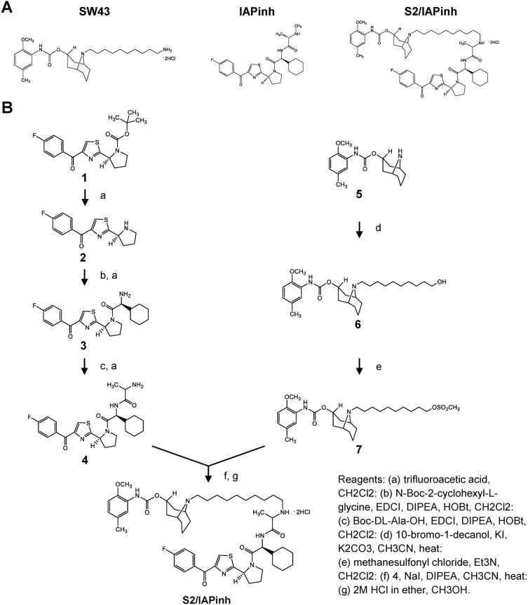
Cancer selective apoptosis remains a therapeutic challenge and off-target toxicity has limited enthusiasm for this target clinically. Sigma-2 ligands (S2) have been shown to enhance the cancer selectivity of small molecule drug candidates by improving internalization. Here, we report the synthesis of a novel drug conjugate, which was created by linking a clinically underperforming SMAC mimetic (second mitochondria-derived activator of caspases; LCL161), an inhibitor (antagonist) of inhibitor of apoptosis proteins (IAPinh) with the sigma-2 ligand SW43, resulting in the new chemical entity S2/IAPinh. Drug potency was assessed via cell viability assays across several pancreatic and ovarian cancer cell lines in comparison with the individual components (S2 and IAPinh) as well as their equimolar mixtures (S2 + IAPinh) both in vitro and in preclinical models of pancreatic and ovarian cancer. Mechanistic studies of S2/IAPinh-mediated cell death were investigated in vitro and in vivo using syngeneic and xenograft mouse models of murine pancreatic and human ovarian cancer, respectively. S2/IAPinh demonstrated markedly improved pharmacological activity in cancer cell lines and primary organoid cultures when compared to the controls. In vivo testing demonstrated a marked reduction in tumor growth rates and increased survival rates when compared to the respective control groups. The predicted mechanism of action of S2/IAPinh was confirmed through assessment of apoptosis pathways and demonstrated strong target degradation (cellular inhibitor of apoptosis proteins-1 [cIAP-1]) and activation of caspases 3 and 8. Taken together, S2/IAPinh demonstrated efficacy in models of pancreatic and ovarian cancer, two challenging malignancies in need of novel treatment concepts. Our data support an in-depth investigation into utilizing S2/IAPinh for the treatment of cancer.


Conflict of interest statement

S.W., W.G.H. and D.S. filed an invention disclosure on the presented technology.

Figures

Figure 1
Figure 1
Compound structures and synthesis schematic of S2/IAPinh. (A) The chemical structures of SW43, IAPinh, and the chemical conjugate S2/IAPinh. (B) Synthesis schematic of S2/IAPinh.
Figure 2
Figure 2
Chemical linkage to sigma-2 ligand SW43 leads to the functionally enhanced drug conjugate S2/IAPinh with increased activity over IAPinh alone. (A) Human pancreatic cancer cell lines AsPC-1, HPAC, and MiaPaCa-2, mouse pancreatic cancer cell lines KP2, and human ovarian cancer cell lines OVCAR3 and OVCAR 8 were treated with different concentrations (0, 3.125, 6.25, 12.5, 25, 50, and 100 µM) of SW43 alone, IAPinh alone, an equimolar mix (SW43 + IAPinh) and S2/IAPinh alone for 24 h and cell viability was determined by CellTiter-Glo Luminescent Viability Assay (Promega). Two-way ANOVA; NS = not significant. (B) Representative images of TUNEL labeled apoptosis cells in HPAC cells treated with vehicle, SW43 (6 µM), IAPinh (6 µM), or S2/IAPinh (6 µM) for 6 h. Nuclei were stained in blue with Hoechst, TUNEL positive cells are in red. Scale bars are equal to 50 µm. (C) Quantification of TUNEL positive cells per area in each treatment group. Data are shown as means ± SEM; **P < 0.01. (D) Organoid cultures from patients with pancreatic cancer (hF3, hF23, hF44, hM1E, hM19A) were treated with escalating concentrations of S2/IAPinh (0, 0.01, 0.02, 0.05, 0.12, 0.28, 0.66, 1.52, 3.6, 8.2, 18.76, 43.2, and 100 µM) for 5 days and cell viability was determined by CellTiter-Glo Luminescent Viability Assay (Promega) (n = 4 per cell line).
Figure 3
Figure 3
Physicochemical stability evaluations of S2/IAPinh. (A) Killing activity of S2/IAPinh (6 µM stock solution) stored at 4 °C, 25 °C, 37 °C, or 60 °C using HPAC reporter cells. An aliquot of the stock solution was frozen immediately (Day 0; black bar, baseline activity). Additional aliquots at the respective temperature condition were removed in 24 h intervals and immediately frozen. At the conclusion of the 7-day experiment, all samples were thawed and subjected to a viability assay on HPAC cells. The day 0 aliquot served as a reference (black bars) with a baseline killing capacity of 77.7% (normalized as dotted lines). (B) Killing activity of S2/IAPinh (6 µM stock solution) following a series of freeze/thaw cycles. An aliquot of the stock solution was frozen immediately (0 freeze/thaw cycles, black bar, baseline activity). Additional aliquots were removed following 5, 10, 20 and 30 freeze/thaw cycles and stored on ice prior to functional evaluation. At the conclusion of the cycling experiment, all samples were thawed and subjected to a viability assay on HPAC cells. The non-cycled aliquot served as a reference (black bar) with a baseline killing capacity of 77.7% (normalized as dotted line).
Figure 4
Figure 4
S2/IAPinh induces activation of the extrinsic apoptosis pathway via degradation of cIAP-1. (A) Schematic diagram of the intrinsic and extrinsic apoptosis pathways: arrows represent stimulation. TNFR, tumor necrosis factor receptor; DR4-5, death receptor 4–5; cIAP-1/2, cellular inhibitor of apoptosis protein-1/2; RIPK1, Receptor-interacting serine/threonine-protein kinase 1. (B) Protein expression of cIAP-1, cIAP-2, and XIAP in HPAC cells treated with vehicle, SW43 (10 µM), IAPinh (10 µM), or S2/IAPinh (10 µM) for 6 h. The precursor and cleaved forms of caspases 3, 8, and 9 were also analyzed for these cells using Wes automated capillary blotting system (Protein Simple). (C) Quantification of protein expression. Relative densitometry of each band normalized to the total protein. Data shown as means ± SEM. **P < 0.01, **** P < 0.0001. (D) Ratio of Caspase 3/7 counts to NucRed counts in HPAC cells treated with vehicle, SW43 (10 µM), IAPinh (10 µM), combination of SW43 (10 µM) and IAPinh (10 µM), or S2/IAPinh (10 µM) measured by the IncuCyte system (Sartorius). Bar graph shows the ratio of Caspase 3/7 at 48 h for each treatment. Data shown as means ± SEM. ****P < 0.001. (E) Activity of cell death in AsPC-1 cells was measured using YOYO-1 iodide on the IncuCyte (Sartorius). Representative images of AsPC-1 cells treated with or without Z-VAD-FMK and S2/IAPinh (10 uM) at baseline and 36 h after treatment. Scale bars are equal to 20 µm. (F) The AUC of lethal fraction at 36 h. Data shown as means ± SEM. ****P < 0.0001.
Figure 5
Figure 5
Antitumoral effect of S2/IAPinh in murine subcutaneous tumor models. (A) KP2 xenograft subcutaneous tumor model using 6-week-old female C57BL/6 mice (12 mice per group) were start treated when the tumor reached approximately 100 mm3. Drug were administered by intraperitoneal injection every day for 21 days with either vehicle (25% cremophor in H2O), 25 µmols/kg/day of SW43 alone, 25 µmols/kg/day of IAPinh alone, combination of 25 µmols/kg/day of SW43 and 25 µmols/kg/day of IAPinh, or 25 µmols/kg/day of S2/IAPinh. (B) Tumor volume of mice during or after each treatment (n = 12/group). There were no significant differences between vehicle, SW43, IAPinh, and combination of SW43 and IAPinh. Data shown as means ± SEM. **** P < 0.0001. (C) Kaplan–Meier survival curve of the mice in each treatment group is shown (n = 12/group). There were no significant differences in survival between vehicle, SW43, IAPinh, and combination of SW43 and IAPinh treated group. **P < 0.01, *** P < 0.001, **** P < 0.0001. (D) Ki-67 staining for tumor samples collected 48 h after each treatment (n = 4/group). Representative images of Ki-67 staining. Nuclei were stained in blue with Hoechst, Ki-67 in green. Scale bars are equal to 20 µm. (E) Quantification of Ki-67 staining per area in each treatment group 48 h after each treatment (n = 4/group). Data are shown as means ± SEM; **P < 0.01, **** P < 0.0001.
Figure 6
Figure 6
S2/IAPinh induces tumor cell death via activation of the extrinsic apoptosis pathway in vivo. KP2 tumor samples collected from animals at 48 h after the treatment with either vehicle, SW43, IAPinh, or S2/IAPinh, were used for analyses. (A) Representative images of TUNEL labeled apoptosis cells in KP2 tumor samples of each group. Nuclei were stained in blue with Hoechst, TUNEL positive cells are in red. Scale bars are equal to 20 µm. (B) Quantification of TUNEL positive cells per area in each group. Data are shown as means ± SEM; ****P < 0.0001. (C) Protein expression of cIAP-1, cIAP-2, and XIAP in KP2 tumor samples of each group. The precursor and cleaved forms of caspases 3, 8, and 9 were also analyzed for these cells using Wes automated capillary blotting system (Protein Simple). (D) Quantification of protein expression. Relative densitometry of each band normalized to the total protein. Data shown as means ± SEM. *P < 0.05, **P < 0.01, **** P < 0.0001.

Similar articles

Cited by

References

    1. Walker JM, et al. Sigma receptors: Biology and function. Pharmacol. Rev. 1990;42:355–402. - PubMed
    1. Quirion R, et al. A proposal for the classification of sigma binding sites. Trends Pharmacol. Sci. 1992;13:85–86. doi: 10.1016/0165-6147(92)90030-A. - DOI - PubMed
    1. Alon A, et al. Identification of the gene that codes for the sigma2 receptor. Proc. Natl. Acad. Sci. U. S. A. 2017;114:7160–7165. doi: 10.1073/pnas.1705154114. - DOI - PMC - PubMed
    1. Riad A, et al. Sigma-2 receptor/TMEM97 and PGRMC-1 increase the rate of internalization of LDL by LDL receptor through the formation of a ternary complex. Sci. Rep. 2018;8:16845. doi: 10.1038/s41598-018-35430-3. - DOI - PMC - PubMed
    1. Wheeler KT, et al. Sigma-2 receptors as a biomarker of proliferation in solid tumours. Br. J. Cancer. 2000;82:1223–1232. doi: 10.1054/bjoc.1999.1067. - DOI - PMC - PubMed