

A comprehensive analysis of CD47 expression in various histological subtypes of soft tissue sarcoma: exploring novel opportunities for macrophage-directed treatments
- PMID: 38493445
- PMCID: PMC10944806
- DOI: 10.1007/s00432-024-05661-1
A comprehensive analysis of CD47 expression in various histological subtypes of soft tissue sarcoma: exploring novel opportunities for macrophage-directed treatments
Abstract
Purpose: The CD47 molecule, often referred to as the "do not eat me" signal, is frequently overexpressed in tumor cells. This signaling pathway limits phagocytosis by macrophages. Our objective was to determine CD47 abundance in various soft tissue sarcomas (STS) to investigate whether it could serve as a potential evasion mechanism for tumor cells. Additionally, we aimed to assess the prognostic value of CD47 expression by examining its association with different clinicopathological factors. This study aimed to elucidate the significance of CD47 in the context of emerging anti-tumor targeting approaches.
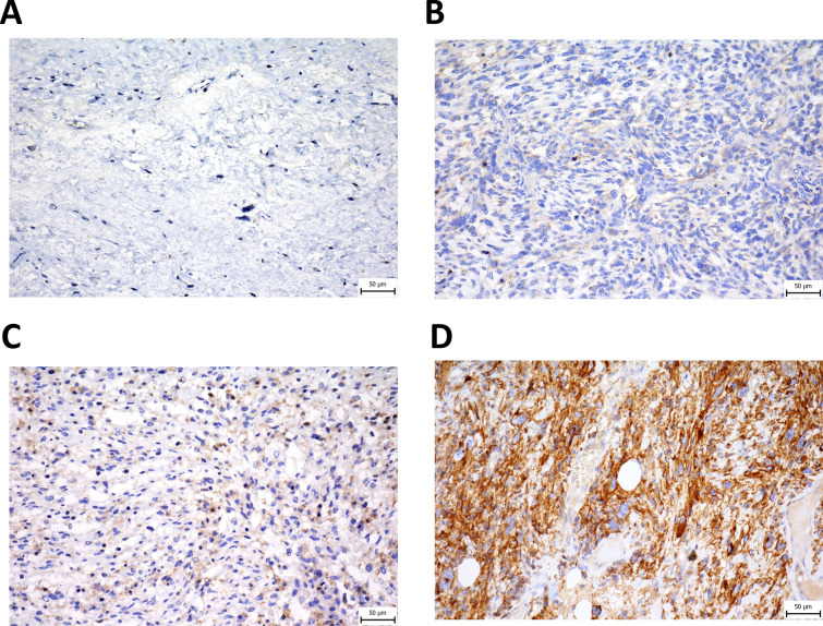
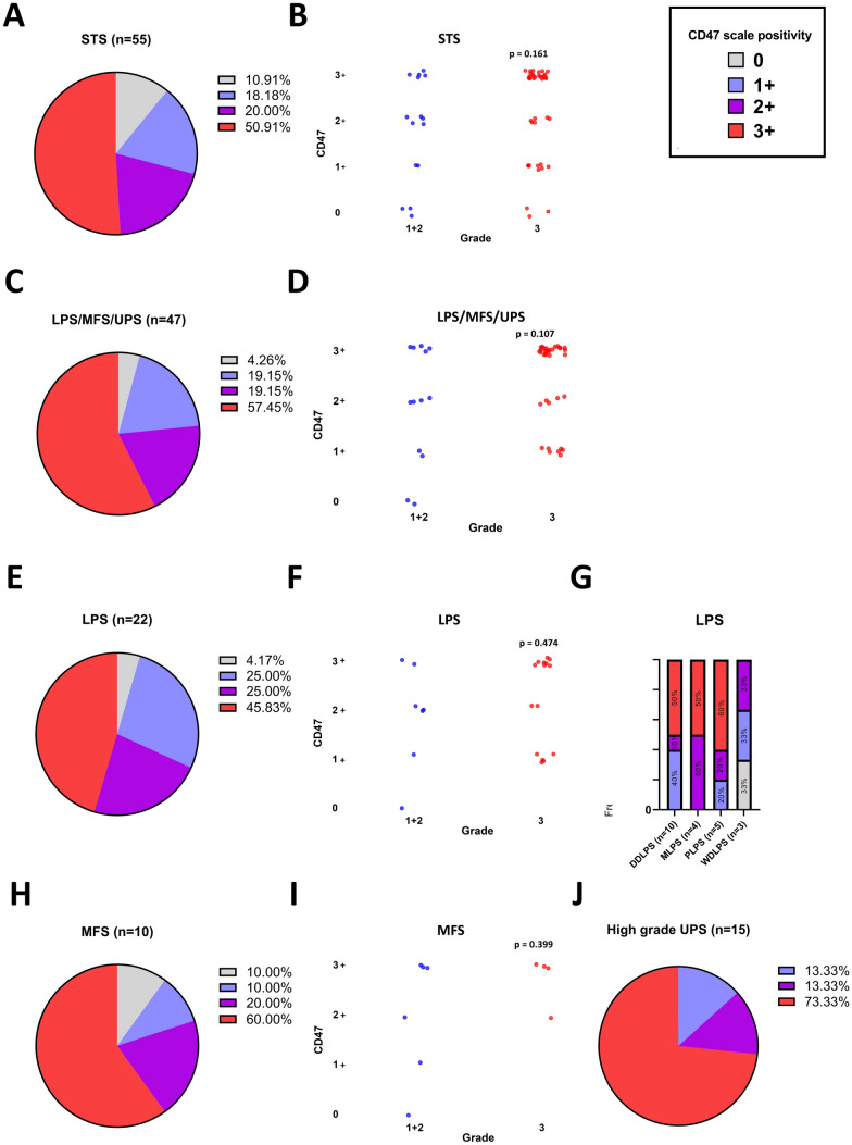
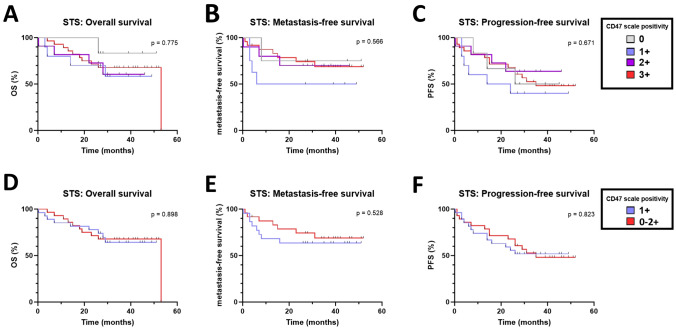
Methods: In this retrospective study, formalin-fixed paraffine-embedded (FFPE) tumor tissues of 55 treatment-naïve patients were evaluated by immunohistochemistry for the abundance of CD47 molecule on tumor cells. The categorization of CD47 positivity was as follows: 0 (no staining of tumor cells), 1 + (less than 1/3 of tumor area positive), 2 + (between 1/3 and 2/3 of tumor area positive), and 3 + (more than 2/3 of tumor area positive for CD47). Next, we compared CD47 abundance between different tumor grades (G1-3). We used Kaplan-Meier survival curves with log-rank test to analyze the differences in survival between patients with different CD47 expression. Moreover, we performed Cox proportional hazards regression model to evaluate the clinical significance of CD47.
Results: CD47 is widely prevalent across distinct STS subtypes. More than 80% of high grade undifferentiated pleiomorphic sarcoma (UPS), 70% of myxofibrosarcoma (MFS) and more than 60% of liposarcoma (LPS) samples displayed a pattern of moderate-to-diffuse positivity. This phenomenon remains consistent regardless of the tumor grade. However, there was a tendency for higher CD47 expression levels in the G3 group compared to the combined G1 + G2 groups when all LPS, MFS, and UPS were analyzed together. No significant associations were observed between CD47 abundance, death, and metastatic status. Additionally, high CD47 expression was associated with a statistically significant increase in progression-free survival in the studied cohort of patients.
Conclusion: This study highlights the potential of the CD47 molecule as a promising immunotherapeutic target in STS, particularly given its elevated expression levels in diverse sarcoma types. Our data showed a notable trend linking CD47 expression to tumor grade, while also suggesting an interesting correlation between enhanced abundance of CD47 expression and a reduced hazard risk of disease progression. Although these findings shed light on different roles of CD47 in STS, further research is crucial to assess its potential in clinical settings.
Keywords: CD47; Immune checkpoint inhibitors; Immunotherapy; Macrophages; Soft tissue sarcoma.
© 2024. The Author(s).
Conflict of interest statement
Iva Benesova, Linda Capkova, Andrej Ozaniak, Pavel Pacas, Katerina Kopeckova, Dominika Galova, Robert Lischke, and Zuzana Ozaniak Strizova declare no conflict of interest. Tomas Buchler: Research support: AstraZeneca, Roche, Bristol Myers Squibb, Exelixis, Merck KGaA, MSD, and Novartis; consulting fees from Bristol Myers Squibb, Astellas, Janssen, and Sanofi/Aventis; payment or honoraria for lectures, presentations, speakers’ bureaus, manuscript writing, or educational events from Ipsen, Bristol-Myers Squibb, AstraZeneca, Roche, Servier, Accord, MSD, and Pfizer. All unrelated to the present paper.
Figures
References
-
- Banks LB, D’Angelo SP (2022) The role of immunotherapy in the management of soft tissue sarcomas: current landscape and future outlook. J Natl Compr Canc Netw 20(7):834–844. 10.6004/jnccn.2022.7027 - PubMed
-
- Chawla SP, Kelly CM, Gordon EM, Quon DV, Moradkhani A, Chua-Alcala VS, Thompson TM, Scheuber A, Bruns I, Allgood VE, Movva S (2022) TTI-621–03: a phase I/II study of TTI-621 in combination with doxorubicin in patients with unresectable or metastatic high-grade leiomyosarcoma (LMS). J Clin Oncol. 10.1200/JCO.2022.40.16_suppl.TPS11593
MeSH terms
Substances
Grants and funding
- GA UK No. 94323/Grantová Agentura, Univerzita Karlova
- AZV NU23J-08-00031/Ministerstvo Zdravotnictví Ceské Republiky
- AZV NU23J-08-00031/Ministerstvo Zdravotnictví Ceské Republiky
- AZV NU23J-08-00031/Ministerstvo Zdravotnictví Ceské Republiky
- Cooperatio Program, Research Area SURG/Univerzita Karlova v Praze
LinkOut - more resources
Full Text Sources
Medical
Research Materials
