. 2024 Jun:406:110109.
doi: 10.1016/j.jneumeth.2024.110109. Epub 2024 Mar 15.

Multimodal active subspace analysis for computing assessment oriented subspaces from neuroimaging data

Affiliations

Multimodal active subspace analysis for computing assessment oriented subspaces from neuroimaging data

Ishaan Batta et al. J Neurosci Methods. 2024 Jun.

Abstract

Background: For successful biomarker discovery, it is essential to develop computational frameworks that summarize high-dimensional neuroimaging data in terms of involved sub-systems of the brain, while also revealing underlying heterogeneous functional and structural changes covarying with specific cognitive and biological traits. However, unsupervised decompositions do not inculcate clinical assessment information, while supervised approaches extract only individual feature importance, thereby impeding qualitative interpretation at the level of subspaces.

New method: We present a novel framework to extract robust multimodal brain subspaces associated with changes in a given cognitive or biological trait. Our approach involves active subspace learning on the gradients of a trained machine learning model followed by clustering to extract and summarize the most salient and consistent subspaces associated with the target variable.

Results: Through a rigorous cross-validation procedure on an Alzheimer's disease (AD) dataset, our framework successfully extracts multimodal subspaces specific to a given clinical assessment (e.g., memory and other cognitive skills), and also retains predictive performance in standard machine learning algorithms. We also show that the salient active subspace directions occur consistently across randomly sub-sampled repetitions of the analysis.

Comparison with existing method(s): Compared to existing unsupervised decompositions based on principle component analysis, the subspace components in our framework retain higher predictive information.

Conclusions: As an important step towards biomarker discovery, our framework not only uncovers AD-related brain regions in the associated brain subspaces, but also enables automated identification of multiple underlying structural and functional sub-systems of the brain that collectively characterize changes in memory and proficiency in cognitive skills related to brain disorders like AD.

Keywords: Brain networks; Functional connectivity; Heterogeneity; Machine learning; Magnetic resonance imaging; Multimodal fusion; Neuroimaging; Subspace analysis.


Conflict of interest statement

Declaration of competing interest Vince Calhoun reports financial support and article publishing charges were provided by the National Institutes of Health. Vince Calhoun reports financial support and article publishing charges were provided by the National Science Foundation. Ishaan Batta, Anees Abrol, and Vince Calhoun report a relationship with Georgia State University that includes: employment. Ishaan Batta, and Vince Calhoun report a relationship with the Georgia Institute of Technology that includes: employment. Vince Calhoun reports a relationship with Emory University that includes: employment.

Figures

Figure 1.
Figure 1.
A schematic diagram showing the step-by-step procedures involved in the overall approach to extract Active Subspace Centers (ASCs) using the Active Subspace Learning (ASL) framework. Starting with fused structural and functional features from brain components as input, ASL is performed for 100 repetitions with support vector regression as the underlying function, followed by the aggregation and clustering of active subspace vectors across repetitions to extract the ASCs. ASCs are multimodal vectors representing consistently occurring important directions in which structural and functional brain features co-vary in association with the target clinical assessments.
Figure 2.
Figure 2.
A total of 53 components corresponding to various brain regions extracted by the scICA procedure using the Neuromark framework are shown in the figure. The components are shown after dividing into 7 brain subdomains (resting state networks), namely Subcortical (SC), Auditory (AUD), Sensorimotor (SM), Visual (VIS), Cognitive Control (CC), Default Mode Network (DMN), and Cerebellar (CB) areas.
Figure 3.
Figure 3.
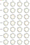
t-SNE embeddings are visualized for the active subspace vectors aggregated from across 100 repetitions of the ASL analysis. K-means clustering was performed on the aggregated vectors for each of the cases of the data from the seven scores and four groupings of the dataset (28 total cases). Top clusters (shown in different random colors) in each case were selected based on the mean fractional contribution (μ>0.1) of the constituent vectors in that cluster (See subsection 2.4). As observable in all cases, the existence of discrete clusters reveals the existence of important directions that are consistently present across multiple repetitions of the analysis on randomly selected subsets of the data. The centroid for each of the discrete clusters is a multimodal vector taken as the active subspace center (ASC), encoding an important direction of collective changes in brain components and connections, which are associated with changes in the target score. (μ: mean fractional contribution, s: mean silhouette score for an individual cluster; s¯: average silhouette score across all clusters, r: correlation between μ and s values of the clusters. Since the correlation (r) values are high in each case, using μ as a metric does not compromise on cluster separation captured by s.)
Figure 4.
Figure 4.
The most contributing multimodal active subspace centers (ASCs), ranked according to the mean fractional contribution (μ) with μ>0.1, computed on all subjects, are shown for the seven scores. Each ASC is represented by a unit norm multimodal direction vector with elements visualized as a multimodal connectogram. The node and edge colors signify the values of the structural and functional elements of the multimodal ASC, respectively. Moreover, it can be noted that the contributive strength for memory-related regions like the hippocampus (Hipp-48, Hipp-83) is negative for scores known for higher values with a decline in memory performance (age, FAQ, CDRSB, ADAS), which is in turn related to shrinkage in these areas. Along similar lines, scores like RAVLT and MMSE, which have a higher value for a better performance, show a positive contribution from the hippocampus in the ASCs. This is in line with the expected contribution of the hippocampus, given its role in memory-related tasks and structural as well as functional changes involved with cognitive decline in Alzheimer’s disease.
Figure 5.
Figure 5.
Top multimodal active subspace centers (ASCs). Each ASC is a unit norm multimodal direction vector with elements visualized as a multimodal connectogram with node and edge colors showing the values of contributive strength of the brain components and connections from the structural and functional parts of the vector. It can be noted that in line with the findings of previous MRI studies on Alzheimer’s disease having greater sensitivity for brain structure compared to function, structural features are the main contributors to the multimodal ASCs corresponding to the AD group, unlike the other groups.
Figure 6.
Figure 6.
Regression performance of ASL features for 100 repetitions of random sub-sampling with 5-fold cross-validation using SVR with the polynomial kernel. The boxplots show the (a) Pearson correlation and (b) normalized root mean squared error (NRMSE) between the predicted and actual values of the 7 clinical scores for the 100 repetitions. Comparison is shown for active subspaces and projections of features using only static functional network connectivity (SFNC), only structural (fSTR), and multimodal input features (fSTRSFNC). It can be noticed that the multimodal features are better at retaining the predictive power of the transformed features than using only structural or only functional features.

Similar articles

References

    1. Abavisani M. and Patel VM (2018). Deep multimodal subspace clustering networks. IEEE Journal of Selected Topics in Signal Processing, 12(6):1601–1614.
    1. Abrol A, Bhattarai M, Fedorov A, Du Y, Plis S, Calhoun V, Initiative ADN, et al. (2020). Deep residual learning for neuroimaging: an application to predict progression to alzheimer’s disease. Journal of neuroscience methods, 339:108701. - PMC - PubMed
    1. Abrol A, Fu Z, Salman M, Silva R, Du Y, Plis S, and Calhoun V. (2021). Deep learning encodes robust discriminative neuroimaging representations to outperform standard machine learning. Nature communications, 12(1):1–17. - PMC - PubMed
    1. Adali T, Akhonda M, and Calhoun VD (2018). Ica and iva for data fusion: An overview and a new approach based on disjoint subspaces. IEEE sensors letters, 3(1):1–4. - PMC - PubMed
    1. Adali T. and Calhoun VD (2022). Reproducibility and replicability in neuroimaging data analysis. Current Opinion in Neurology, 35(4):475–481. - PMC - PubMed

Publication types