

A conserved asparagine residue stabilizes iron binding in Manduca sexta transferrin-1
- PMID: 38494145
- PMCID: PMC11018507
- DOI: 10.1016/j.ibmb.2024.104109
A conserved asparagine residue stabilizes iron binding in Manduca sexta transferrin-1
Abstract
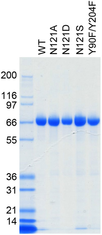
Transferrin 1 (Tsf1) is an insect-specific iron-binding protein that is abundant in hemolymph and other extracellular fluids. It binds iron tightly at neutral pH and releases iron under acidic conditions. Tsf1 influences the distribution of iron in the body and protects against infection. Elucidating the mechanisms by which Tsf1 achieves these functions will require an understanding of how Tsf1 binds and releases iron. Previously, crystallized Tsf1 from Manduca sexta was shown to have a novel type of iron coordination that involves four iron-binding ligands: two tyrosine residues (Tyr90 and Tyr204), a buried carbonate anion, and a solvent-exposed carbonate anion. The solvent-exposed carbonate anion was bound by a single amino acid residue, a highly conserved asparagine at position 121 (Asn121); thus, we predicted that Asn121 would be essential for high-affinity iron binding. To test this hypothesis, we analyzed the iron-binding and -release properties of five forms of recombinant Tsf1: wild-type, a Y90F/Y204F double mutant (negative control), and three Asn121 mutants (N121A, N121D and N121S). Each of the Asn121 mutants exhibited altered spectral properties, confirming that Asn121 contributes to iron coordination. The N121D and N121S mutations resulted in slightly lower affinity for iron, especially at acidic pH, while iron binding and release by the N121A mutant was indistinguishable from that of the wild-type protein. The surprisingly minor consequences of mutating Asn121, despite its high degree of conservation in diverse insect species, suggest that Asn121 may play a role that is essential in vivo but non-essential for high affinity iron binding in vitro.
Keywords: Carbonate; Hemolymph; Insect; Iron; Mutant; Transferrin.
Copyright © 2024 Elsevier Ltd. All rights reserved.
Conflict of interest statement
Declarations of interest: none
Figures


Similar articles
-
Structural insight into the novel iron-coordination and domain interactions of transferrin-1 from a model insect, Manduca sexta.Protein Sci. 2021 Feb;30(2):408-422. doi: 10.1002/pro.3999. Epub 2020 Nov 28. Protein Sci. 2021. PMID: 33197096 Free PMC article.
-
Iron binding and release properties of transferrin-1 from Drosophila melanogaster and Manduca sexta: Implications for insect iron homeostasis.Insect Biochem Mol Biol. 2020 Oct;125:103438. doi: 10.1016/j.ibmb.2020.103438. Epub 2020 Jul 29. Insect Biochem Mol Biol. 2020. PMID: 32735914 Free PMC article.
-
Phenotypic analyses, protein localization, and bacteriostatic activity of Drosophila melanogaster transferrin-1.Insect Biochem Mol Biol. 2022 Aug;147:103811. doi: 10.1016/j.ibmb.2022.103811. Epub 2022 Jul 1. Insect Biochem Mol Biol. 2022. PMID: 35781032 Free PMC article.
-
Anion binding properties of the transferrins. Implications for function.Biochim Biophys Acta. 2012 Mar;1820(3):348-61. doi: 10.1016/j.bbagen.2011.07.017. Epub 2011 Aug 5. Biochim Biophys Acta. 2012. PMID: 21846492 Review.
-
Insect transferrins: multifunctional proteins.Biochim Biophys Acta. 2012 Mar;1820(3):437-51. doi: 10.1016/j.bbagen.2011.07.011. Epub 2011 Jul 23. Biochim Biophys Acta. 2012. PMID: 21810453 Review.
References
MeSH terms
Substances
Grants and funding
LinkOut - more resources
Full Text Sources
