. 2024 Mar 17;81(1):144.
doi: 10.1007/s00018-024-05174-7.

Molecular engineering of a spheroid-penetrating phage nanovector for photodynamic treatment of colon cancer cells

Affiliations

Molecular engineering of a spheroid-penetrating phage nanovector for photodynamic treatment of colon cancer cells

Eleonora Turrini et al. Cell Mol Life Sci. .

Abstract

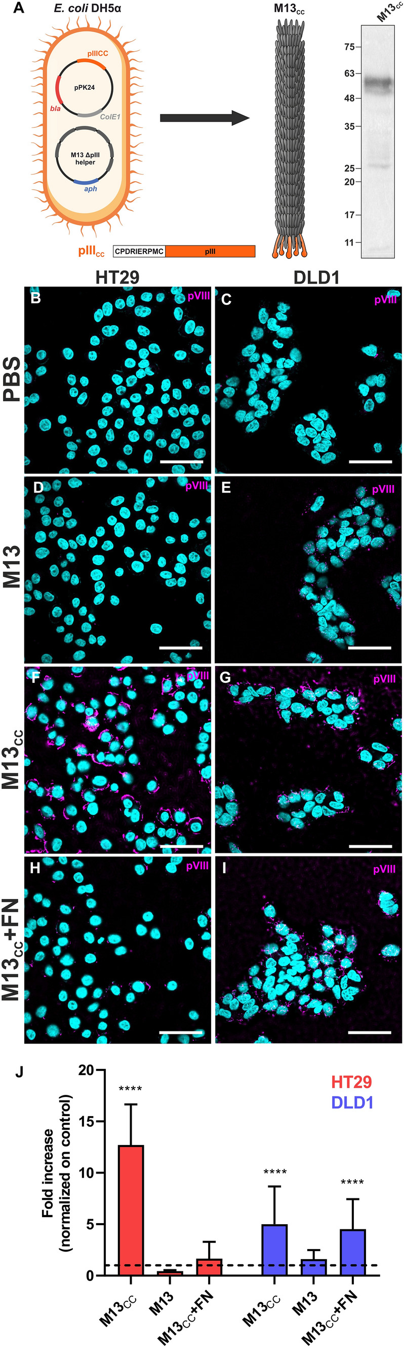
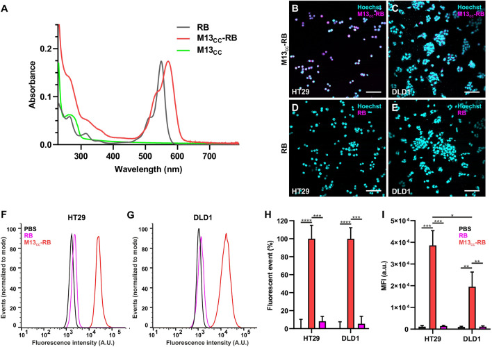
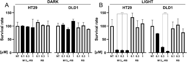
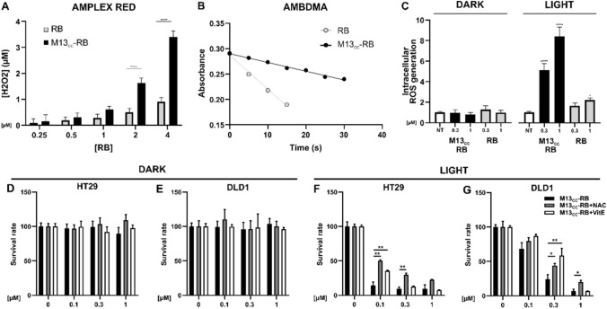
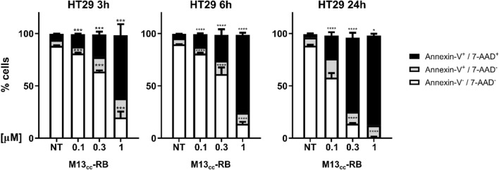
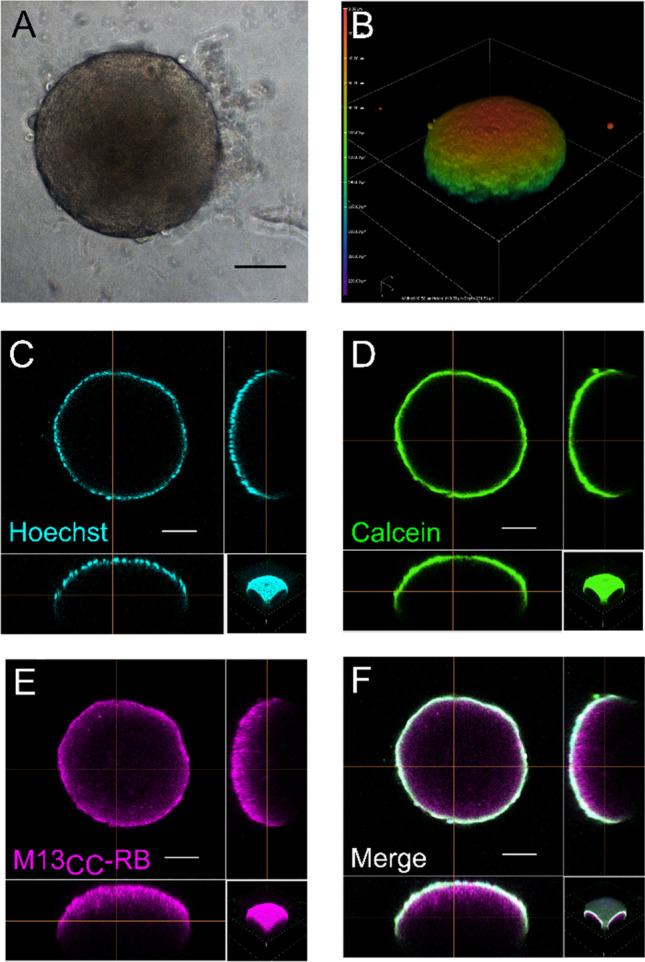
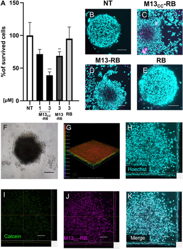
Photodynamic therapy (PDT) represents an emerging strategy to treat various malignancies, including colorectal cancer (CC), the third most common cancer type. This work presents an engineered M13 phage retargeted towards CC cells through pentavalent display of a disulfide-constrained peptide nonamer. The M13CC nanovector was conjugated with the photosensitizer Rose Bengal (RB), and the photodynamic anticancer effects of the resulting M13CC-RB bioconjugate were investigated on CC cells. We show that upon irradiation M13CC-RB is able to impair CC cell viability, and that this effect depends on i) photosensitizer concentration and ii) targeting efficiency towards CC cell lines, proving the specificity of the vector compared to unmodified M13 phage. We also demonstrate that M13CC-RB enhances generation and intracellular accumulation of reactive oxygen species (ROS) triggering CC cell death. To further investigate the anticancer potential of M13CC-RB, we performed PDT experiments on 3D CC spheroids, proving, for the first time, the ability of engineered M13 phage conjugates to deeply penetrate multicellular spheroids. Moreover, significant photodynamic effects, including spheroid disruption and cytotoxicity, were readily triggered at picomolar concentrations of the phage vector. Taken together, our results promote engineered M13 phages as promising nanovector platform for targeted photosensitization, paving the way to novel adjuvant approaches to fight CC malignancies.

Keywords: Bacteriophage; Colorectal cancer; M13; Nanovector; PDT; Spheroids.


Conflict of interest statement

The authors have no relevant financial or non-financial interests to disclose.

Figures

Fig. 1
Fig. 1
M13CC targets CC cells. A Phage modification scheme, The CPDIRIERPMC coding sequence was cloned in frame with pIII (orange) generating the pPK24 plasmid. Transformed bacteria superinfected with Hyperphage helper produce modified phages. Immunoblot of M13CC demonstrates incorporation of modified pIII in the purified virions. Immunohistochemical confocal microscopy of HT29 (B, D, F, H) and DLD1 (C, E, G, I) cell lines incubated with PBS (B, C), M13 (D, E), M13CC (F, G) and M13CC after preincubation with fibronectin (FN) (H, I). Nuclei are in cyan while the major coating protein pVIII of phage is in magenta. Scale bar = 50 µm. J Quantitative analysis performed on confocal images. Fluorescence intensity detected in confocal images were expressed as fold increase in comparison to the control PBS (dashed line) (n = 20). Statistical significance was calculated by one-way parametric ANOVA in comparison to the control (PBS); ****p < 0.0001
Fig. 2
Fig. 2
Enhanced targeting of the M13CC-RB towards CC cells, significantly higher for HT29 cells. A UV–VIS absorption spectra of RB (grey), M13CC-RB (red) and M13CC (green). (B) Microscopic analysis of the retargeting properties of the engineered phage 1 µM RB equivalents tested on B HT29 and C DLD1 CC cell lines. Microscopic analyses were performed also with free RB on D HT29 and E DLD1. Flow cytometry analysis of the retargeting ability of either free RB (pink) or M13CC -RB (red) on F HT29 and G DLD1, PBS was used as control (in black). H Percentage of fluorescent events calculated via flow cytometry. I Mean fluorescence intensity of HT29 cells and DLD1 cells incubated with free RB (in pink), M13CC-RB (in red) and PBS (in black). All graphs and calculations were performed using FlowJo™. Statistical significance was calculated by one-way parametric ANOVA followed by Dunnet’s multiple comparison test * p < 0.05; *** p < 0.001; **** p < 0.0001; n = 3
Fig. 3
Fig. 3
Photoirradiated M13CC-RB strongly decreases CC cell viability. Cytotoxic effects of M13CC-RB or RB alone on HT29 and DLD1 cells kept in the dark (DARK, A) or irradiated for 30 min with a white led bulb (LIGHT, B), 24 h after treatment. The same sensitizer concentrations (RB equivalents) were used to compare M13CC-RB and RB alone. Cell viability was analyzed using MTT test on three independent biological replicates (n = 3). Statistical significance was calculated by one-way parametric ANOVA followed by Dunnet’s multiple comparison test; * p < 0.05; *** p < 0.001; **** p < 0.0001 compared to untreated cells (NT)
Fig. 4
Fig. 4
M13CC-RB increases ROS intracellular levels after irradiation. A Quantification of peroxides, assayed by AR test, produced by different concentrations of RB (white squares) and M13CC-RB (black squares) after 30 min of white LED irradiation. B Singlet oxygen generation produced by RB (grey dots) and M13CC-RB (black dots) at increasing irradiation times, monitored by the decrease of UV–Vis absorption of ABMDMA. C Intracellular ROS generation measured with ROS-Glo assay (Promega) after exposure to M13CC-RB in presence or absence (dark) of irradiation (30 min) of HT29 cells on three independent biological replicates (n = 3). Statistical significance was calculated by two-way ANOVA followed by Dunnet’s multiple comparisons test. * p < 0.05; **** p < 0.0001 compared to untreated cells (NT). PDT with M13CC-RB in presence of ROS inhibitor was evaluated on D,F HT29 and E,G DLD1 cells. Cells were treated with M13CC-RB in presence of PBS (black bars), NAC (grey bars) or vitamin E (VitE, white bars) and then D,E kept in dark conditions or F,G irradiated. Statistical significance was calculated by one-way parametric ANOVA followed by Dunnet’s multiple comparisons test; * p < 0.05; ** p < 0.01; (n = 3)
Fig. 5
Fig. 5
Cell death mechanisms induced by M13CC-RB mediated photosensitization. Percentage of Annexin-V/7-AAD, Annexin-V+/7-AAD, Annexin-V+/7-AAD+ HT29 cells after 3, 6 or 24 h from treatment with photoactivated M13CC-RB on three independent biological replicates (n = 3). Statistical significance was calculated by two-way Anova followed by Dunnet’s multiple comparisons test. * p < 0.05; *** p < 0.001; **** p < 0.0001 compared to untreated cells (NT)
Fig. 6
Fig. 6
M13CC-RB deeply penetrates CC multicellular spheroids. Evaluation of multicellular spheroid conformation in A bright field and B confocal microscopy after 24 h from incubation with M13CC-RB at 3 µM concentration of PS. The heatmap highlights the Z-depth coding, showing the relative height of cells in the confocal stack, demonstrating the integrity of the CC spheroid. Spheroid penetration of M13CC-RB: C Hoechst, cyan; D Calcein AM, green; E M13CC-RB, magenta; F merge. Scale bars = 100 µm
Fig. 7
Fig. 7
M13CC-RB impairs CC multicellular spheroid structure and viability upon irradiation. A Cytotoxic effects of M13CC-RB (1 µM and 3 µM), M13-RB (3 µM) or RB alone (3 µM) after 30 min of irradiation. 3D CellTiter-Glo® was used as viability assay on three independent biological replicates (n = 3). Statistical significance was calculated by one-way ANOVA followed by Dunnet’s multiple comparison test; ** p < 0.01; *** p < 0.001 compared to untreated cells (NT). B Untreated control spheroid; C spheroid disaggregation after M13CC-RB-mediated PDT; D partial spheroid disaggregation after PDT with M13-RB; E lack of spheroid disaggregation after PDT with RB alone. Evaluation of multicellular spheroid conformation in F bright field and G confocal microscopy after PDT with M13CC-RB at 3 µM concentration of PS. The heatmap highlights the Z-depth coding, showing the relative height of cells in the confocal stack, and demonstrating loss of spheroid integrity. Spheroid disaggregation and loss of viability after M13CC-RB photoactivation: H Hoechst, cyan; I Calcein AM, green; J M13CC-RB, magenta; K merge. Scale bars = 100 µm

Similar articles

Cited by

References

    1. IARC 2022 https://www.iarc.who.int/featured-news/colorectal-cancer-awareness-month...
    1. Hodgkinson N, Kruger CA, Abrahamse H. Targeted photodynamic therapy as potential treatment modality for the eradication of colon cancer and colon cancer stem cells. Tumour Biol J Int Soc Oncodevelopmental Biol Med. 2017;39:1010428317734691. doi: 10.1177/1010428317734691. - DOI - PubMed
    1. Shi H, Sadler PJ. How promising is phototherapy for cancer? Br J Cancer. 2020;123:871–873. doi: 10.1038/s41416-020-0926-3. - DOI - PMC - PubMed
    1. Mroz P, Yaroslavsky A, Kharkwal GB, Hamblin MR. Cell death pathways in photodynamic therapy of cancer. Cancers. 2011;3:2516–2539. doi: 10.3390/cancers3022516. - DOI - PMC - PubMed
    1. Niculescu A-G, Grumezescu AM. Photodynamic therapy—an up-to-date review. Appl Sci. 2021;11:3626. doi: 10.3390/app11083626. - DOI

LinkOut - more resources