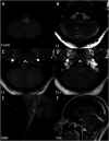
Listeria monocytogenes brain abscesses presenting as contiguous, tubular rim-enhancing lesions on Magnetic Resonance Imaging: Case series and literature review
- PMID: 38494758
- PMCID: PMC11571425
- DOI: 10.1177/19714009241240054
Listeria monocytogenes brain abscesses presenting as contiguous, tubular rim-enhancing lesions on Magnetic Resonance Imaging: Case series and literature review
Abstract
Listeriosis has more than a 50% mortality when the central nervous system is involved, necessitating rapid diagnosis and treatment. We present four patients with brain abscesses in the setting of diagnosed neurolisteriosis, all of which demonstrated an odd presentation of multiple small, contiguous tubular lesions with rim enhancement on magnetic resonance imaging. Our review of published cases of neurolisteriosis suggests that this may be a useful pattern to identify neurolisteriosis abscesses, allowing earlier detection and therapy.
Keywords: Listeria brain abscess; Listeria monocytogenes; Magnetic Resonance Imaging; infectious imaging sign; neurolisteriosis.
Conflict of interest statement
Declaration of conflicting interestsThe author(s) declared no potential conflicts of interest with respect to the research, authorship, and/or publication of this article.
Figures


Similar articles
-
Multiple cerebral abscesses because of Listeria monocytogenes: three case reports and a literature review of supratentorial listerial brain abscess(es).Surg Neurol. 2003 Apr;59(4):320-8. doi: 10.1016/s0090-3019(03)00056-9. Surg Neurol. 2003. PMID: 12748019 Review.
-
Worm-like appearance of Listeria monocytogenes brain abscess: presentation of three cases.Neuroradiology. 2020 Sep;62(9):1189-1193. doi: 10.1007/s00234-020-02441-9. Epub 2020 May 14. Neuroradiology. 2020. PMID: 32405729
-
Multiple cortical brain abscesses due to Listeria monocytogenes in an immunocompetent patient.Trop Doct. 2018 Apr;48(2):160-163. doi: 10.1177/0049475517728670. Epub 2017 Aug 29. Trop Doct. 2018. PMID: 28849735
-
Spreading of multiple Listeria monocytogenes abscesses via central nervous system fiber tracts: case report.J Neurosurg. 2015 Dec;123(6):1593-9. doi: 10.3171/2014.12.JNS142100. Epub 2015 Jun 19. J Neurosurg. 2015. PMID: 26090836
-
Cerebellar abscess caused by Listeria monocytogenes in a liver transplant patient.Transpl Infect Dis. 2013 Dec;15(6):E224-8. doi: 10.1111/tid.12145. Epub 2013 Oct 23. Transpl Infect Dis. 2013. PMID: 24298984 Review.
References
-
- Zhang C, Yi Z. Brain abscess caused by Listeria monocytogenes: a case report and literature review. Ann Palliat Med 2022; 11: 3356. - PubMed
-
- Bokhari MR, Mesfin FB. Brain abscess. In: StatPearls. Treasure Island (FL): StatPearls Publishing, 2023, https://www.ncbi.nlm.nih.gov/books/NBK441841/(accessed 31 August 2023). - PubMed
Publication types
MeSH terms
LinkOut - more resources
Full Text Sources
Medical

