. 2024 May 25;56(5):776-788.
doi: 10.3724/abbs.2024027.

Maslinic acid alleviates intervertebral disc degeneration by inhibiting the PI3K/AKT and NF-κB signaling pathways

Affiliations

Maslinic acid alleviates intervertebral disc degeneration by inhibiting the PI3K/AKT and NF-κB signaling pathways

Yichen Que et al. Acta Biochim Biophys Sin (Shanghai). .

Abstract

Intervertebral disc degeneration (IDD) is the cause of low back pain (LBP), and recent research has suggested that inflammatory cytokines play a significant role in this process. Maslinic acid (MA), a natural compound found in olive plants ( Olea europaea), has anti-inflammatory properties, but its potential for treating IDD is unclear. The current study aims to investigate the effects of MA on TNFα-induced IDD in vitro and in other in vivo models. Our findings suggest that MA ameliorates the imbalance of the extracellular matrix (ECM) and mitigates senescence by upregulating aggrecan and collagen II levels as well as downregulating MMP and ADAMTS levels in nucleus pulposus cells (NPCs). It can also impede the progression of IDD in rats. We further find that MA significantly affects the PI3K/AKT and NF-κB pathways in TNFα-induced NPCs determined by RNA-seq and experimental verification, while the AKT agonist Sc-79 eliminates these signaling cascades. Furthermore, molecular docking simulation shows that MA directly binds to PI3K. Dysfunction of the PI3K/AKT pathway and ECM metabolism has also been confirmed in clinical specimens of degenerated nucleus pulposus. This study demonstrates that MA may hold promise as a therapeutic agent for alleviating ECM metabolism disorders and senescence to treat IDD.

Keywords: NF-κB; PI3K; intervertebral disc degeneration; maslinic acid; senescence.


Conflict of interest statement

The authors declare that they have no conflict of interest.

Figures

None
Figure 1
The impact of MA on ECM anabolism and catabolism in TNFα-treated NPCs (A) Chemical architecture of MA. (B) The cytotoxicity of MA to NPCs was evaluated at designated time points by CCK-8 assay at various concentrations. (C–H) The relative mRNA levels of COL2A1, ACAN, ADAMTS5, ADAMTS4, MMP13 and MMP9 were quantified via qPCR. (I) The protein expression levels of ACAN, COL2A1, ADAMTS4, MMP9, ADAMTS5 and MMP13 were assessed in NPCs treated with or without MA in the presence of TNFα. *P<0.05, **P<0.01, ***P<0.001.
None
Figure 2
MA mitigated TNFα-induced senescence in NPCs (A‒D) The expressions of COL2A1, ACAN, MMP13, and ADAMTS4 were detected through immunofluorescence staining for nuclear visualization. Scale bar: 20 μm. (E) Fluorescence was quantified using ImageJ software. (F) Cellular senescence in TNFα-treated NPCs was assessed through β-gal staining. Scale bar: 50 μm. (G) Quantitative analysis of the percentage of senescent NPCs expressing β-gal in (F). (H) The protein expression levels of senescence markers (MMP3, p21 and p16) were assessed. ***P<0.001.
None
Figure 3
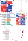
MA modulated the activation of the NF-κB and PI3K/AKT signaling pathways in TNFα-stimulated NPCs (A) Heatmap displaying the DEGs identified by RNA-seq in NPCs treated with TNFα alone or in combination with MA. (B) Map of the gene distribution in relation to volcanic activity. (C) The GO analysis terms, which included three analyses: BP (biological process), CC (cellular component), and MF (molecular function). (D) KEGG pathway enrichment analysis revealed pathways. (E) Heatmap displaying the expression levels of genes, including ACAN, MMPs, ADAMTSs, and inflammatory factors.
None
Figure 4
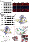
MA treatment modulated the activation of the NF-κB and PI3K/AKT signaling pathways in TNFα-stimulated NPCs (A) Western blot analysis of the NF-κB pathway in TNFα-treated NPCs with or without MA. (B) Immunofluorescence was utilized to detect p65. Scale bar: 20 μm. (C) Western blot analysis of the PI3K pathway in TNFα-treated NPCs treated with or without MA. (D) Ribbon model depicting the structure of PI3K. (E,F) The space-filling model illustrates the interaction between MA and PI3K, which results in a reaction with an interaction energy of –10.35 kcal mol−1.
None
Figure 5
The AKT agonist Sc-79 effectively counteracted the inhibitory effects of MA on the PI3K/AKT/NF-κB signaling pathway (A) Western blot analysis of the PI3K/AKT/NF-κB pathway was performed after 30 min of treatment, as indicated. (B) Immunofluorescence staining was performed to determine the nuclear translocation of NF-κB p65. Scale bar: 20 μm. (C) Cellular senescence was assessed through β-gal staining. Scale bar: 50 μm. (D) Quantitative analysis of the percentage of senescent NPCs. (E) Western blot analysis was performed to detect senescence markers (MMP3, p21 and p16). (F) The protein expression levels of ACAN, COL2A1, ADAMTS4, ADAMTS5 and MMP13 were assessed via western blot analysis. (G–J) Immunofluorescence staining was utilized to detect the expressions of COL2A1, ACACN, MMP13 and ADAMTS4. Scale bar: 20 μm. (K) Fluorescence intensity was quantified using ImageJ. ***P<0.001.
None
Figure 6
MA mitigates IDD progression in a rat model (A) MRI of rat tails with corresponding treatments. Scale bar: 5 mm. (B) Analysis of Pfirrmann scores from images across multiple groups (n=5). (C,D) Illustrations depicting H&E staining and cross-SO staining of samples from various experimental groups. (E–G) Immunohistochemical staining was performed to detect the expressions of COL2A1, ACAN, MMP13, p21, p-PI3K and NF-κB p65 in rat intervertebral discs. Scale bar: 100 μm. (H–M) Statistical analysis of the immunohistochemical staining results. *P<0.05, **P<0.01, ***P<0.001.

Similar articles

Cited by

References

    1. Hartvigsen J, Hancock MJ, Kongsted A, Louw Q, Ferreira ML, Genevay S, Hoy D, et al. What low back pain is and why we need to pay attention. Lancet. . 2018;391:2356–2367. doi: 10.1016/S0140-6736(18)30480-X. - DOI - PubMed
    1. Dieleman JL, Cao J, Chapin A, Chen C, Li Z, Liu A, Horst C, et al. US health care spending by payer and health condition, 1996–2016. JAMA. . 2020;323:863–884. doi: 10.1001/jama.2020.0734. - DOI - PMC - PubMed
    1. Lyu FJ, Cui H, Pan H, MC Cheung K, Cao X, Iatridis JC, Zheng Z. Painful intervertebral disc degeneration and inflammation: from laboratory evidence to clinical interventions. Bone Res. . 2021;9:7. doi: 10.1038/s41413-020-00125-x. - DOI - PMC - PubMed
    1. Wang F, Cai F, Shi R, Wang XH, Wu XT. Aging and age related stresses: a senescence mechanism of intervertebral disc degeneration. Osteoarthritis Cartilage. . 2016;24:398–408. doi: 10.1016/j.joca.2015.09.019. - DOI - PubMed
    1. Qiu X, Liang T, Wu Z, Zhu Y, Gao W, Gao B, Qiu J, et al. Melatonin reverses tumor necrosis factor-alpha-induced metabolic disturbance of human nucleus pulposus cells via MTNR1B/Gαi2/YAP signaling. Int J Biol Sci. . 2022;18:2202–2219. doi: 10.7150/ijbs.65973. - DOI - PMC - PubMed

MeSH terms