. 2024 Mar 1:15:1346756.
doi: 10.3389/fphar.2024.1346756. eCollection 2024.

Targeted intracellular delivery of dimeric STINGa by two pHLIP peptides for treatment of solid tumors

Affiliations

Targeted intracellular delivery of dimeric STINGa by two pHLIP peptides for treatment of solid tumors

Anna Moshnikova et al. Front Pharmacol. .

Abstract

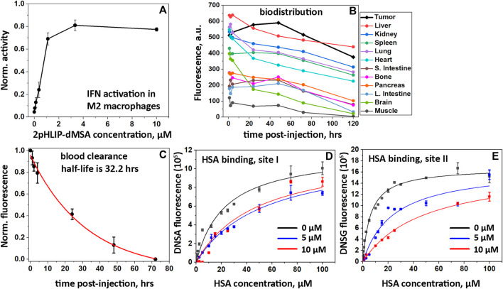
Introduction: We have developed a delivery approach that uses two pHLIP peptides that collaborate in the targeted intracellular delivery of a single payload, dimeric STINGa (dMSA). Methods: dMSA was conjugated with two pHLIP peptides via S-S cleavable self-immolating linkers to form 2pHLIP-dMSA. Results: Biophysical studies were carried out to confirm pH-triggered interactions of the 2pHLIP-dMSA with membrane lipid bilayers. The kinetics of linker self-immolation and dMSA release, the pharmacokinetics, the binding to plasma proteins, the stability of the agent in plasma, the targeting and resulting cytokine activation in tumors, and the biodistribution of the construct was investigated. This is the first study demonstrating that combining the energy of the membrane-associated folding of two pHLIPs can be utilized to enhance the targeted intracellular delivery of large therapeutic cargo payloads. Discussion: Linking two pHLIPs to the cargo extends blood half-life, and targeted delivery of dimeric STINGa induces tumor eradication and the development of robust anti-cancer immunity.

Keywords: biophysics; cold tumors; imaging; immuno-suppressive tumors; tumor acidity.


Conflict of interest statement

DE, OA, and YR are founders of pHLIP, Inc., and they have shares in the company. pHLIP, Inc. sponsored synthesis and purification of dMSA and modified dMSA prepared by the Iris Biotech GmbH. The remaining authors declare that the research was conducted in the absence of any commercial or financial relationships that could be construed as a potential conflict of interest.

Figures

FIGURE 1
FIGURE 1
Schematic representation of 2pHLIP-dMSA interaction with a membrane lipid bilayer. The chemical structure of dMSA and proposed interaction with 2 pHLIP peptides are shown (A). In (B), schematics are shown of 2pHLIP-dMSA reversible interactions with the membranes of cells with normal surface pH (pH = 7.2) in healthy tissue and membranes of acidic cells with surface pH < 6.2 found in diseased tissues.
FIGURE 2
FIGURE 2
Interactions of 2pHLIP-dMSA with liposome lipid bilayers. Fluorescence (A) and CD (B) spectra of 2pHLIP-dMSA in solution at pH 7.4 (black lines), in the presence of POPC liposomes at pH 7.4 (blue lines) and in the presence of POPC liposomes at pH 3.5 (red lines) are shown. CD spectral signals were normalized per peptide and multiplied by (−1), since the D-amino acid pHLIP peptides used in the study invert the usual CD spectrum. pH-dependent transitions monitored by changes of CD spectral signal in presence of POPC liposomes (experimental points and fitting curves, red, with 95% confidence interval, pink) are presented (C). Kinetics of fluorescence changes triggered by pH drops (D, E) and pH increases (F) in presence of POPC liposomes are shown.
FIGURE 3
FIGURE 3
In vitro IFN activations, biodistribution, PK and HSA binding. Activation of the IFN signaling pathway induced by 2pHLIP-dMSA in THP1-Blue-ISG cells polarized by PMA, IL-4/IL-13 into M2-type macrophages is shown (A). The results were normalized to the activity of dMSA alone at the maximum concentration tested, which was taken as 1. Kinetics of 2(ICG-pHLIP)-dMSA targeting CT26 tumor and clearance of the agent from major organs are shown (B). The mean fluorescence per area was calculated for each organ and tissue collected at different timepoints after single RO injection of 2(ICG-pHLIP)-dMSA (50 μM 100 μL). Normalized fluorescence recorded in blood, which was collected at different time points after single RO injection of 2(ICG-pHLIP)-dMSA is shown (mean and Sd.) (C). The data were fitted with an exponential function (red line). Changes of DNSA (D) and DNSG (E) fluorescence with increasing HSA concentration in absence (black symbols) or presence of 5 µM (blue symbols) or 10 µM (red symbols) of 2pHLIP-dMSA. The global fitting was performed with each set of data (D and E) and the fitting curves are shown as black, blue and red lines.
FIGURE 4
FIGURE 4
Activation of cytokines. Levels of TNF-α (A and B), IL-6 (C and D), INF-β (E and F), IFN-γ (G and H) and INF-α (I and J) cytokines are shown in tumors and serum as established by ELISA at 6 and 24 h after a single RO injection of 2pHLIP-dMSA (300 μM 500 µL) in comparison to control mice (all points, mean, and SE are shown, * indicates that the p-level is < 0.02 and ** indicates that the p-level is < 0.002, otherwise differences are statistically insignificant, p-levels were calculated using the Kolmogorov-Smirnov two-tailed nonparametric test).
FIGURE 5
FIGURE 5
Eradication of CT26 tumors and development of immune memory. The experimental design is presented (A). Right flank CT26 tumor growth curves in Balb/c mice are shown after two IP injections of 2pHLIP-dMSA (B), dMSA (D) at dose 300 μM 500 μL per injection, 2pHLIP-dMSA at lower dose (L.d.) 100 μM 500 μL per injection (C), and vehicle (E) performed on days 1 and 3, when tumors had reached about 100–150 mm3 in volume. The surviving mice were re-injected with CT26 cancer cells in the contralateral left flank on day 65, indicated by red triangle. The average changes of mouse body weights after two IP injections of 2pHLIP-dMSA (F) and 2pHLIP-dMSA at lower dose (G) are shown. Kaplan-Meier survival plots are shown (H).

References

    1. Chamma H., Vila I. K., Taffoni C., Turtoi A., Laguette N. (2022). Activation of STING in the pancreatic tumor microenvironment: a novel therapeutic opportunity. Cancer Lett. 538, 215694. 10.1016/j.canlet.2022.215694 - DOI - PubMed
    1. Cheng C. J., Bahal R., Babar I. A., Pincus Z., Barrera F., Liu C., et al. (2015). MicroRNA silencing for cancer therapy targeted to the tumour microenvironment. Nature 518, 107–110. 10.1038/nature13905 - DOI - PMC - PubMed
    1. Crawford T., Moshnikova A., Roles S., Weerakkody D., DuPont M., Carter L. M., et al. (2020). pHLIP ICG for delineation of tumors and blood flow during fluorescence-guided surgery. Sci. Rep. 10, 18356. 10.1038/s41598-020-75443-5 - DOI - PMC - PubMed
    1. Dane E. L., Belessiotis-Richards A., Backlund C., Wang J., Hidaka K., Milling L. E., et al. (2022). STING agonist delivery by tumour-penetrating PEG-lipid nanodiscs primes robust anticancer immunity. Nat. Mater 21, 710–720. 10.1038/s41563-022-01251-z - DOI - PMC - PubMed
    1. Decout A., Katz J. D., Venkatraman S., Ablasser A. (2021). The cGAS-STING pathway as a therapeutic target in inflammatory diseases. Nat. Rev. Immunol. 21, 548–569. 10.1038/s41577-021-00524-z - DOI - PMC - PubMed

LinkOut - more resources