. 2024 Mar 5;10(5):e27400.
doi: 10.1016/j.heliyon.2024.e27400. eCollection 2024 Mar 15.

Mycobacterium tuberculosis Rv2617c is involved in stress response and phage infection resistance

Affiliations

Mycobacterium tuberculosis Rv2617c is involved in stress response and phage infection resistance

Liadrine Moukendza Koundi et al. Heliyon. .

Abstract

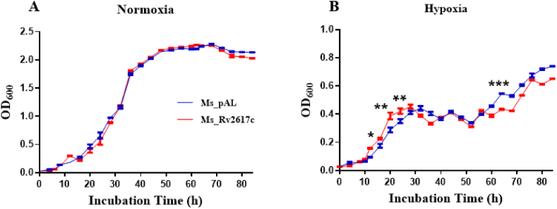
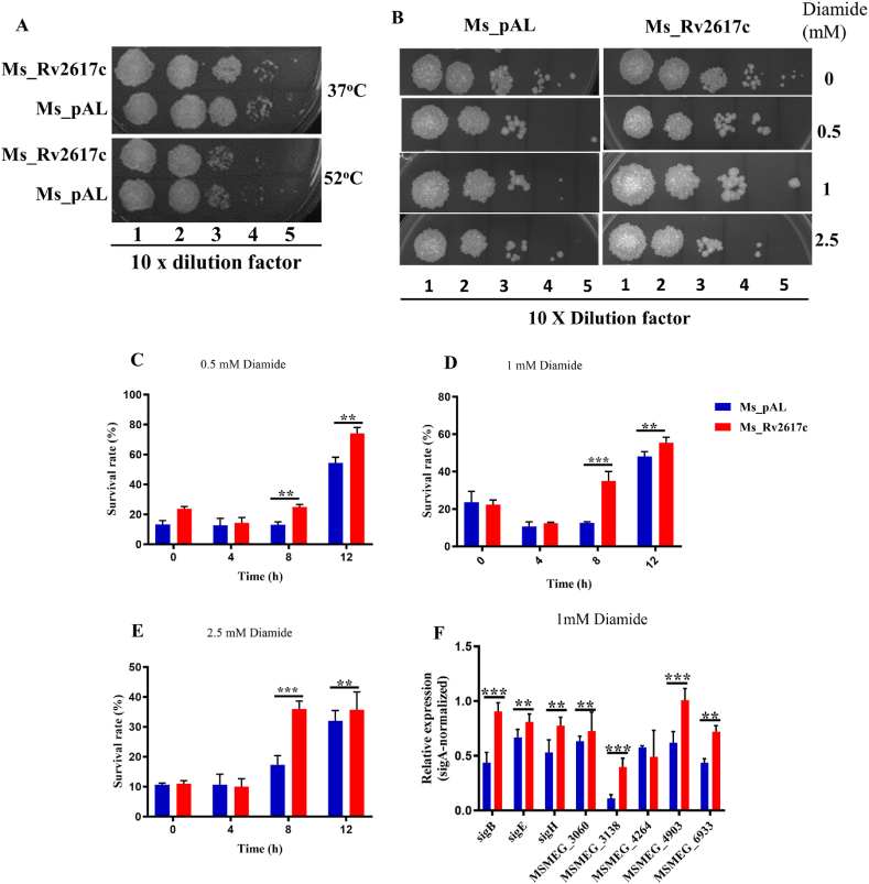
Mycobacterium tuberculosis (M. tuberculosis) is the pathogen of human tuberculosis (TB). Resistance to numerous in vivo stresses, including oxidative stress, is determinant for M. tuberculosis intracellular survival, and understanding associated mechanisms is crucial for developing new therapeutic strategies. M. tuberculosis Rv2617c has been associated with oxidative stress response when interacting with other proteins in M. tuberculosis; however, its functional promiscuity and underlying molecular mechanisms remain elusive. In this study, we investigated the phenotypic changes of Mycobacterium smegmatis (M. smegmatis) expressing Rv2617c (Ms_Rv2617c) and its behavior in the presence of various in vitro stresses and phage infections. We found that Rv2617c conferred resistance to SDS and diamide while sensitizing M. smegmatis to oxidative stress (H2O2) and altered mycobacterial phenotypic properties (single-cell clone and motility), suggestive of reprogrammed mycobacterial cell wall lipid contents exemplified by increased cell wall permeability. Interestingly, we also found that Rv2617c promoted M. smegmatis resistance to infection by phages (SWU1, SWU2, D29, and TM4) and kept phage TM4 from destroying mycobacterial biofilms. Our findings provide new insights into the role of Rv2617c in resistance to oxide and acid stresses and report for the first time on its role in phage resistance in Mycobacterium.

Keywords: DoxX domain; Mycobacteriophage resistance; Mycobacterium tuberculosis; Rv2617c; Stress responses; Virulence.


Conflict of interest statement

The authors declare that they have no known competing financial interests or personal relationships that could have appeared to influence the work reported in this paper.

Figures

Fig. 1
Fig. 1
Rv2617c protein is highly conserved among mycobacteria. (A) A phylogenetic tree was obtained after blasting the M. tuberculosis Rv2617c protein sequence in NCBI. M. tuberculosis H37Rv Rv2617c is closer to its orthologs in M. bovis, M. tuberculosis 1418, M. decipiens, and M. gordonae. (B) Multiple sequence alignment (MSA) of M. tuberculosis Rv2617c protein with its orthologs from M. bovis, M. tuberculosis 1418, M. decipiens, and M. gordonae. The conserved DoxX domain is shaded in red, and the AF_AFI6XER9F1_Chain1 is the predicted secondary structure model of the probable transmembrane protein (Rv2617c) from M. tuberculosis DoxX family proteins (https://www.rcsb.org/structure/AF_AFI6XER9F1).
Fig. 2
Fig. 2
Heterologous expression of M. tuberculosis Rv2617c in M. smegmatis. (A) PCR amplification of Rv2617c from M. tuberculosis H37Rv genome. (B) The constructed Ms_Rv2617c strain was cultured in a 7H9 medium supplemented with acetamide and then subjected to PCR amplification to detect the Rv2617c gene with specific primers. (C) The cell lysates prepared from acetamide-induced Ms_pAL and Ms_Rv2617c cultures were analyzed by SDS-PAGE and visualized under UV after Coomassie Brilliant Blue staining to identify the His-Tag-associated Rv2617c protein. (D) Western Blot analysis of cell wall lysates of both strains. The His-tag-associated Rv2617c protein was targeted with anti-His-tag antibodies. See original and uncropped images in supplementary material.
Fig. 3
Fig. 3
Bacterial growth assessment. (A) Ms_pAL and Ms_Rv2617c were grown in MB 7H9 medium supplemented with 0.05% (v/v) Tween80, 0.5% glycerol, and 1% acetamide. (B) Discoloration of methylene blue (0.2 μg/mL) around 10 h after inoculation of bacteria in flasks sealed with hypoxia plugs indicated the onset of hypoxia. The OD600 was determined at an interval of 4 h, and the data are the mean of three replicates. Data are shown as the means ± SD of three independent experiments (*p < 0.05, **p < 0.01, and ***p < 0.001).
Fig. 4
Fig. 4
Rv2617c is involved in mycobacterial stress response. (A) Disc diffusion assay of 1% H2O2 on agar plates with 0.7% agar mixed with Ms_pAL or Ms_Rv2617c, and (B) the diameter of the complete inhibition zone was measured after 3 days of incubation at 37 °C. (C) Disc diffusion assay of 5% SDS on agar plates with 0.7% agar mixed with Ms_pAL or Ms_Rv2617c, and (D) the diameter of the complete inhibition zone was measured after 3 days of incubation at 37 °C. (E), (F), and (G) Ms_pAL and Ms_Rv2617c were exposed to adjusted 7H9 medium pHs ( 4.5, 5.5, and 6.5) for 3 h, 6 h, and 9 h, respectively. Ms_Rv2617c showed significant growth defects, after 6 h and 9 h incubation in pH 5.5-adjusted 7H9 medium compared to Ms_pAL. All cultures have been previously induced with 1% acetamide and data are shown as the means ± SD of 3 independent experiments (*p < 0.05, **p < 0.01, and ***p < 0.001).
Fig. 5
Fig. 5
Rv2617c is involved in diamide stress resistance. (A) Survival of Ms_pAL and Ms_Rv2617c after treatment under heat shock conditions. Aliquots from heated (37 °C and 52 °C) samples were 10-fold serially diluted and spotted on 7H10 plates. B) Bacterial samples (OD600 = 0.5) were 10-fold serially diluted and spotted on 7H10 plates containing indicated concentrations of diamide. (C), (D), and (E) Ms_pAL and Ms_Rv2617c were exposed to 0.5, 1, and 2.5 mM diamide for 4 h, 8 h, and 12 h, respectively. The results were analyzed after incubation at 37 °C for 3 days. (F) RT-PCR analysis of the expression levels of critical genes of known mycobacterial stress regulators. All cultures have been previously induced with 1% acetamide, and data are shown as the means ± SD of 3 independent experiments (*p < 0.05, **p < 0.01, and ***p < 0.001).
Fig. 6
Fig. 6
Uptake of decreasing concentrations of EtBr (8–0.25 μg/mL) and Nile Red (160–5 μM). (A) Ms_Rv2617c's accumulation of EtBr is higher than that of Ms_pAL. (B) Ms_Rv2617c's accumulation of Nile Red is lower than that of Ms_pAL. The data reported represent the means ± SD of 2 independent experiments (*p < 0.05, **p < 0.01, and ***p < 0.001).
Fig. 7
Fig. 7
Rv2617c is not involved in antibiotic susceptibility. 10-fold serial dilutions of Ms_pAL and Ms_Rv2617c cultures (OD600 = 0.5, ∼4*108 cells/mL) were spotted on MB 7H10 without or with nalidixic acid (20 μg/mL), norfloxacin (1 μg/mL), ofloxacin (0.1 μg/mL), pyrazinamide (25 μg/mL), rifampicin (4 μg/mL), or chloramphenicol (16 μg/mL). The results were recorded after 3 days of incubation at 37 °C.
Fig. 8
Fig. 8
Rv2617c overexpression affects M. smegmatis phenotypes. (A) Biofilm formation of Ms_pAL and Ms_Rv2617c in acetamide-induced 7H9 medium. (B) Single-cell colony of strains on MB 7H10 plates. Few Ms_Rv2617c colonies are observed on the plate and show a less compact shape in the center. (C) Sliding motility assay of Ms_pAL and Ms_Rv2617c on 0.3% 7H9 agar. Ms_Rv2617c increases in size. Induced bacterial cultures (OD600 = 0.5, ∼4*108) were used for the above assays. (D) Sizes of colonies on the culture dishes were measured after 3 days of incubation at 37 °C. The Figure scale is indicated where required. Data are shown as the means ± SD of 3 independent experiments (*p < 0.05, **p < 0.01, and ***p < 0.001).
Fig. 9
Fig. 9
Phage susceptibility of Ms_pAL and Ms_Rv2617c. Spot clearance of lawns of Ms_pAL and Ms_Rv2617c cultures (OD600 = 1, ∼8*108 cells/mL) after treatment with 10-fold diluted (10−1 to 10−8) mycobacteriophages SWU1, SWU2, D29, TM4, and L5. The results were compared after 3 days of incubation at 37 °C.
Fig. 10
Fig. 10
Bacterial growth kinetics upon phage infections. (A), (B), (C), (D), and (E) Ms_pAL and Ms_Rv2617c strains were grown with mycobacteriophages SWU1, SWU2, D29, TM4, and L5, respectively. OD600 was measured every 4 h during 88 h. The black arrows indicate mycobacteriophage inoculation time in mycobacterial cultures. Data are shown as the means ± SD of 3 independent experiments (*p < 0.05, **p < 0.01, ***p < 0.001, and ****p < 0.001).
Fig. 11
Fig. 11
Rv2617c promotes mycobacterial biofilm formation in the presence of phages. Ms_pAL And Ms_Rv2617c strains were cultured in the presence or absence of SWU1, SWU2, D29, TM4, and L5 phages (titers 10−9 PFU/mL), respectively. After 3 days of incubation at 37 °C, the results were retrieved and compared.

Similar articles

References

    1. Bo H., et al. Mycobacterium tuberculosis-macrophage interaction: molecular updates. Front. Cell. Infect. Microbiol. 2023;13 - PMC - PubMed
    1. Yu X., et al. Mycobacterium tuberculosis PE_PGRS1 promotes mycobacteria intracellular survival via reducing the concentration of intracellular free Ca(2+) and suppressing endoplasmic reticulum stress. Mol. Immunol. 2023;154:24–32. - PubMed
    1. Shariq M., et al. The exploitation of host autophagy and ubiquitin machinery by Mycobacterium tuberculosis in shaping immune responses and host defense during infection. Autophagy. 2023;19(1):3–23. - PMC - PubMed
    1. Bhattacharyya A., et al. Oxidative stress: an essential factor in the pathogenesis of gastrointestinal mucosal diseases. Physiol. Rev. 2014;94(2):329–354. - PMC - PubMed
    1. Forrellad M.A., et al. Rv2617c and P36 are virulence factors of pathogenic mycobacteria involved in resistance to oxidative stress. Virulence. 2019;10(1):1026–1033. - PMC - PubMed

LinkOut - more resources