A rare case of resection of a mucinous cystic neoplasm originating from the extrahepatic bile duct with cholangioscopic imaging
- PMID: 38495468
- PMCID: PMC10941567
- DOI: 10.1002/deo2.349
A rare case of resection of a mucinous cystic neoplasm originating from the extrahepatic bile duct with cholangioscopic imaging
Abstract
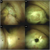
A 29-year-old woman was admitted to our hospital for examination of obstructive jaundice and an extrahepatic bile duct lesion. Contrast-enhanced computed tomography revealed a 20 mm cystic lesion with a thin external capsule in the common hepatic duct. Cholangioscopy revealed translucent oval masses with capillary vessels attached to the bile duct walls. The surface was mostly smooth yet partially irregular with redness, suggesting that the masses were epithelial neoplasms. Histological findings of cholangioscopy-guided targeted biopsies of the mass showed subepithelial spindle cell proliferation with no atypical epithelium. The patient underwent an extrahepatic bile duct resection to confirm the pathological diagnosis. Immunohistochemistry of surgical specimens revealed that the spindle cells were positive for estrogen and progesterone receptors. Finally, the cystic lesion with ovarian-like stroma was diagnosed as a mucinous cystic neoplasm with low-grade intraepithelial neoplasia. This is the first report of cholangioscopic imaging of a biliary mucinous cyctic neoplasm. Cholangioscopic imaging can be helpful in the differential diagnosis of biliary neoplasms and in the determination of treatment strategies.
Keywords: biliary cystadenoma; cholangioscopic imaging; extrahepatic bile duct; mucinous cystic neoplasm; obstructive jaundice.
© 2024 The Authors. DEN Open published by John Wiley & Sons Australia, Ltd on behalf of Japan Gastroenterological Endoscopy Society.
Conflict of interest statement
None.
Figures


References
-
- Zen Y, Pedica F, Patcha VR et al. Mucinous cystic neoplasms of the liver: A clinicopathological study and comparison with intraductal papillary neoplasms of the bile duct. Mod Pathol 2011; 24: 1079–1089. - PubMed
-
- Kulpatcharapong S, Pittayanon R, Kerr SJ, Rerknimitr R. Diagnostic performance of digital and video cholangioscopes in patients with suspected malignant biliary strictures: A systematic review and meta‐analysis. Surg Endosc 2022; 36: 2827–2841. - PubMed
-
- Basturk O, Klimstra DS, Nakamura Y et al. Mucinous cystic neoplasms of the liver and biliary system. In: WHO Classification of Tumours Editorial Board, (ed). WHO Classification of Tumours of the Digestive System, 5th edn. Lyon: International Agency for Research on Cancer, 2019; 250–253.
-
- Tanaka M, Fernández‐del Castillo C, Adsay V et al. International Consensus Guidelines 2012 for the management of IPMN and MCN of the pancreas. Pancreatology 2012; 12: 183–197. - PubMed
Publication types
LinkOut - more resources
Full Text Sources
