

3D polarization-interference holographic histology for wavelet-based differentiation of the polycrystalline component of biological tissues with different necrotic states. Forensic applications
- PMID: 38495527
- PMCID: PMC10943250
- DOI: 10.1117/1.JBO.29.5.052920
3D polarization-interference holographic histology for wavelet-based differentiation of the polycrystalline component of biological tissues with different necrotic states. Forensic applications
Abstract
Significance: The interference-holographic method of phase scanning of fields of scattered laser radiation is proposed. The effectiveness of this method for the selection of variously dispersed components is demonstrated. This method made it possible to obtain polarization maps of biological tissues at a high level of depolarized background. The scale-selective analysis of such maps was used to determine necrotic changes in the optically anisotropic architectonics of biological tissues.
Objective: Development and experimental approbation of layered phase polarimetry of repeatedly scattered fields in diffuse layers of biological tissues. Application of scale-selective processing of the found coordinate distributions of polarization states in various phase sections of object fields. Determination of criteria (markers) for histological differential diagnosis of the causes of necrotic changes in optical anisotropy of biological tissues.
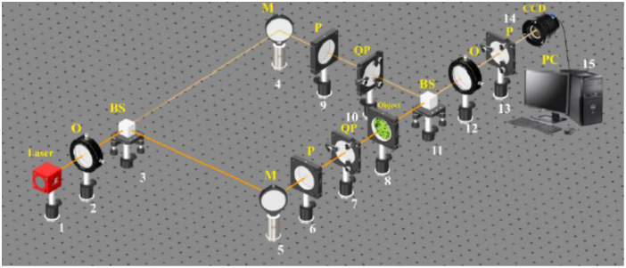
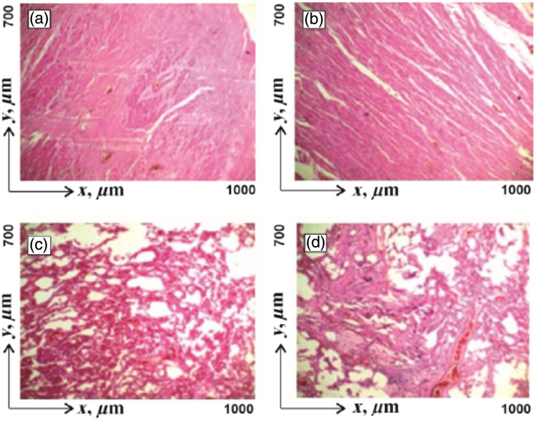
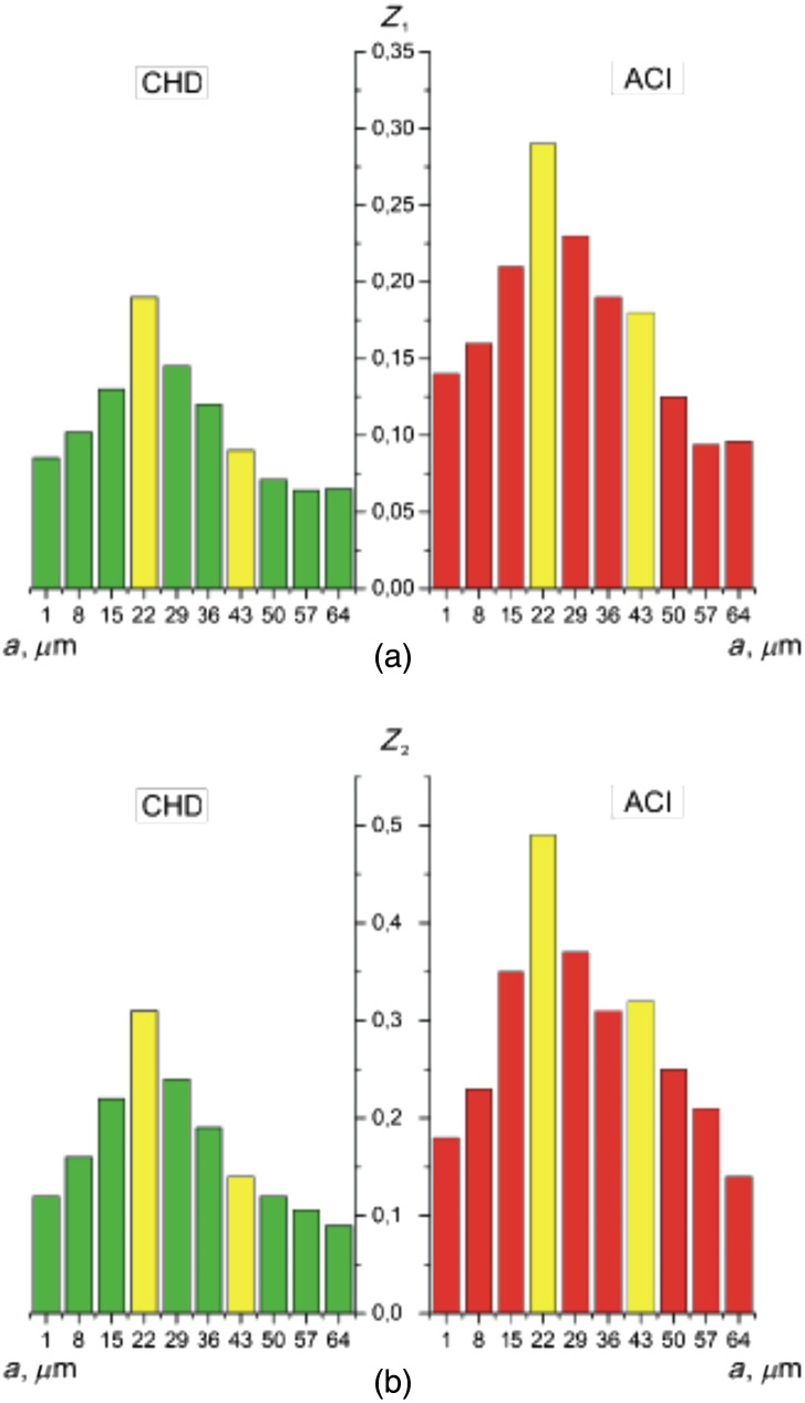
Approach: We used a synthesis of three instrumental and analytical methods. Polarization-interference registration of laser radiation scattered by a sample of biological tissue. Digital holographic reconstruction and layered phase scanning of distributions of complex amplitudes of the object field. Analytical determination of polarization maps of various phase cross-sections of repeatedly scattered radiation. Application of wavelet analysis of the distributions of polarization states in the phase plane of a single scattered component of an object field. Determination of criteria (markers) for differential diagnosis of necrotic changes in biological tissues with different morphological structure. Two cases are considered. The first case is the myocardium of those who died as a result of coronary heart disease and acute coronary insufficiency. The second case is lung tissue samples of deceased with bronchial asthma and fibrosis.
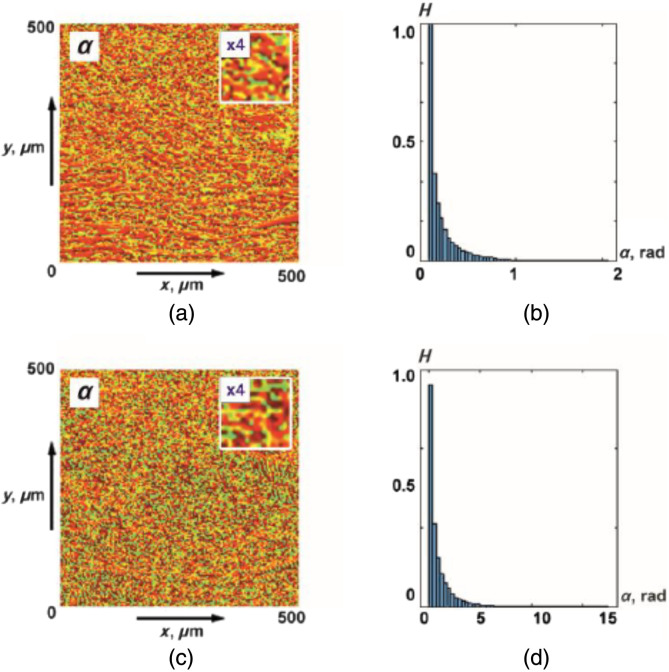
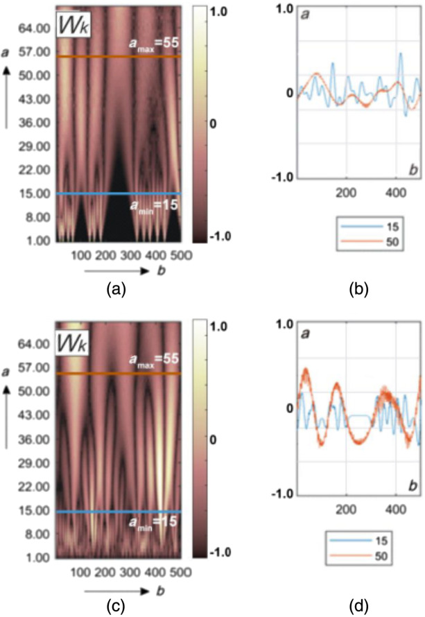
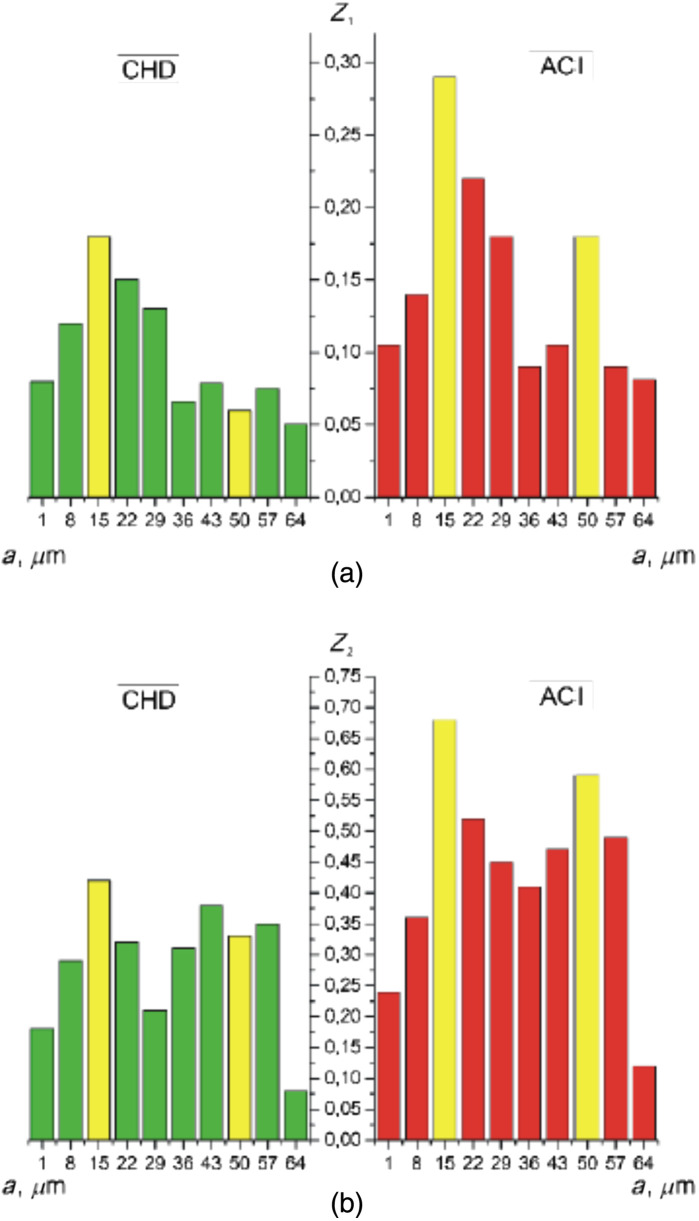
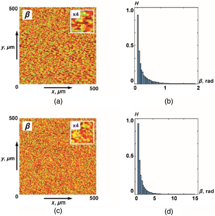
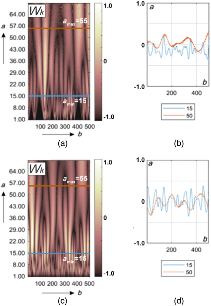
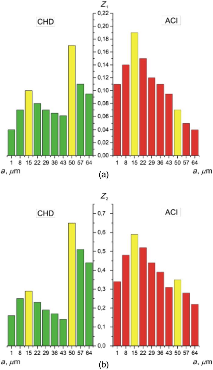
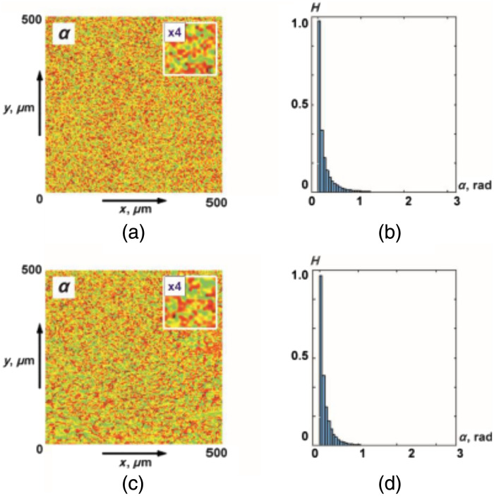
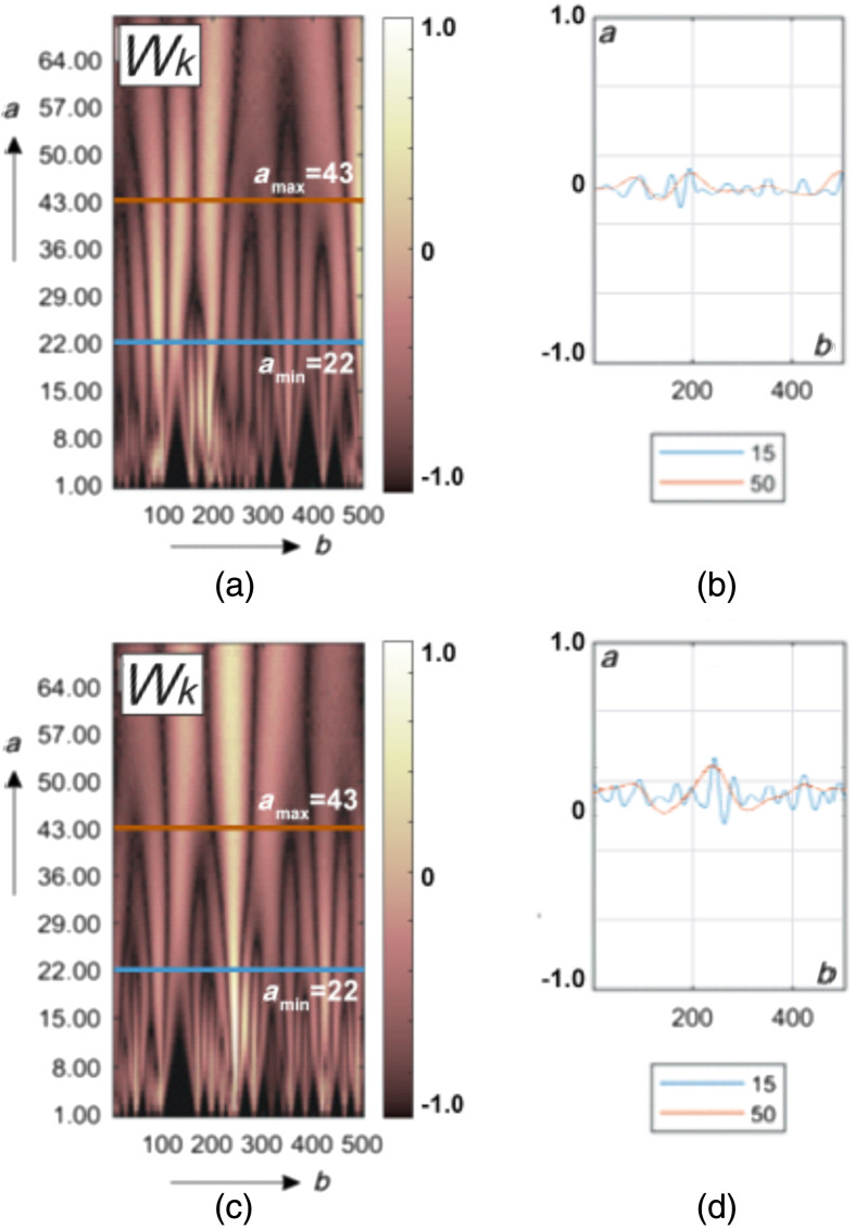
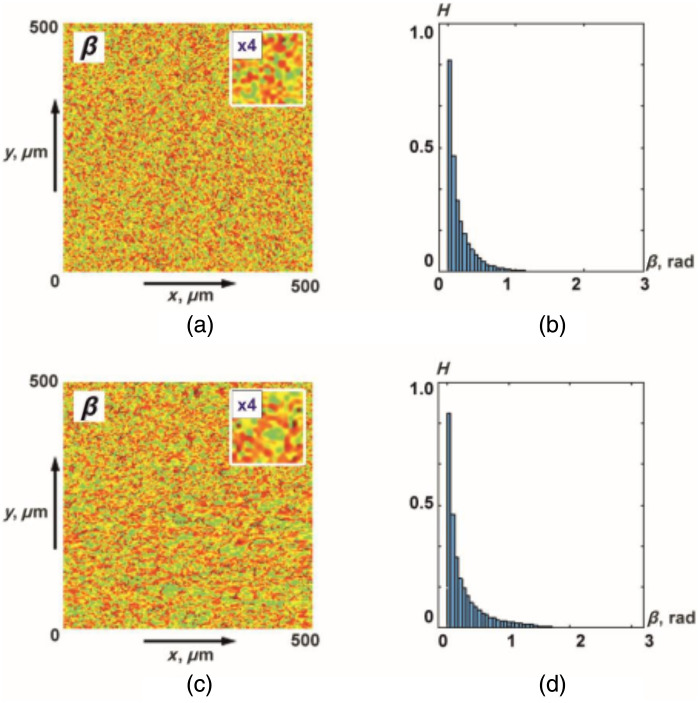
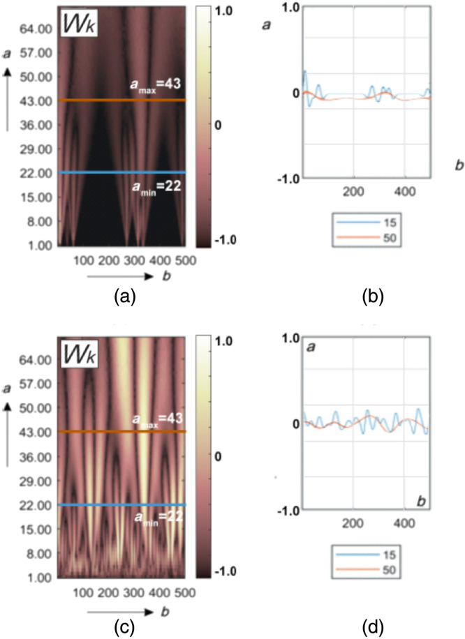
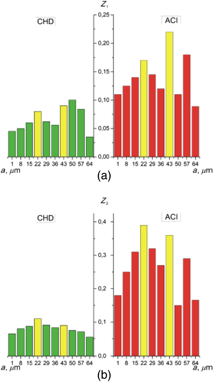
Results: A method of polarization-interference mapping of diffuse object fields of biological tissues has been developed and experimentally implemented. With the help of digital holographic reconstruction of the distributions of complex amplitudes, polarization maps in various phase sections of a diffuse object field are found. The wavelet analysis of azimuth and ellipticity distributions of polarization in the phase plane of a single scattered component of laser radiation is used. Scenarios for changing the amplitude of the wavelet coefficients for different scales of the scanning salt-like MHAT function are determined. Statistical moments of the first to fourth orders are determined for the distributions of the amplitudes of the wavelet coefficients of the azimuth maps and the ellipticity of polarization. As a result, diagnostic markers of necrotic changes in the myocardium and lung tissue were determined. The statistical criteria found are the basis for determining the accuracy of their differential diagnosis of various necrotic states of biological tissues.
Conclusions: Necrotic changes caused by "coronary artery disease-acute coronary insufficiency" and "asthma-pulmonary fibrosis" were demonstrated by the method of wavelet differentiation with polarization interference with excellent accuracy.
Keywords: biological tissue; holography; interference; lungs tissue; myocardium; optical anisotropy; polarization; statistical moments; wavelet analysis.
© 2024 The Authors.
Figures
Similar articles
-
3D digital polarization-holographic wavelet histology in determining the duration of mechanical damage to the myocardium.J Biophotonics. 2024 Mar;17(3):e202300372. doi: 10.1002/jbio.202300372. Epub 2024 Jan 10. J Biophotonics. 2024. PMID: 37915304
-
Mueller-Matrix Interferometric Multifractal Scaling of Optically Anisotropic Architectonics of Diffuse Blood Facies: Fundamental and Applied Aspects.J Biophotonics. 2025 Mar;18(3):e202400412. doi: 10.1002/jbio.202400412. Epub 2025 Jan 6. J Biophotonics. 2025. PMID: 39757863
-
Differential Mueller matrix imaging of partially depolarizing optically anisotropic biological tissues.Lasers Med Sci. 2020 Jun;35(4):877-891. doi: 10.1007/s10103-019-02878-2. Epub 2019 Nov 20. Lasers Med Sci. 2020. PMID: 31749042 Free PMC article.
-
Embossed topographic depolarisation maps of biological tissues with different morphological structures.Sci Rep. 2021 Feb 16;11(1):3871. doi: 10.1038/s41598-021-83017-2. Sci Rep. 2021. PMID: 33594107 Free PMC article.
-
A Review of Polarization-Sensitive Materials for Polarization Holography.Materials (Basel). 2020 Dec 6;13(23):5562. doi: 10.3390/ma13235562. Materials (Basel). 2020. PMID: 33291278 Free PMC article. Review.
References
-
- Tuchin V. V., “Light scattering study of tissues,” Phys.-Usp. 40(5), 495 (1997).PHUSEY10.1070/PU1997v040n05ABEH000236 - DOI
-
- Jacques S., “Polarized light imaging of biological tissues,” in Handbook of Biomedical Optics, Boas D. A., Pitris C., Ramanujam N., Eds., pp. 649–669, CRC Press, Boca Raton, Florida: (2011).
-
- Ushenko A. G., Pishak V. P., “Laser polarimetry of biological tissues: principles and applications,” in Handbook of Coherent Domain Optical Methods, Tuchin V. V., Ed., pp. 93–138, Springer US; (2004).
Publication types
MeSH terms
LinkOut - more resources
Full Text Sources
Research Materials
