. 2024 Mar 1:6:1346781.
doi: 10.3389/fgeed.2024.1346781. eCollection 2024.

Nuclease-free precise genome editing corrects MECP2 mutations associated with Rett syndrome

Affiliations

Nuclease-free precise genome editing corrects MECP2 mutations associated with Rett syndrome

Swati Bijlani et al. Front Genome Ed. .

Abstract

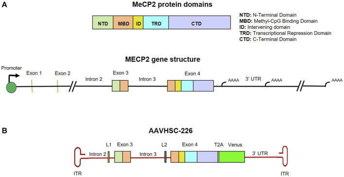
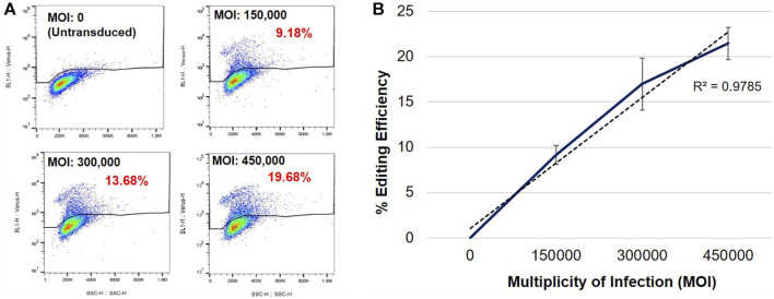
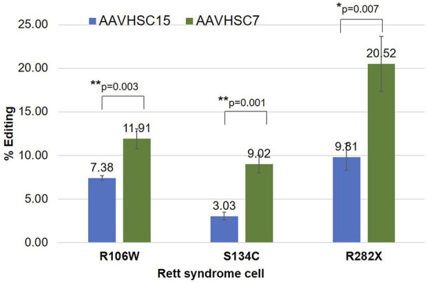
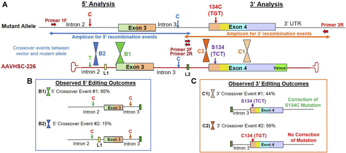
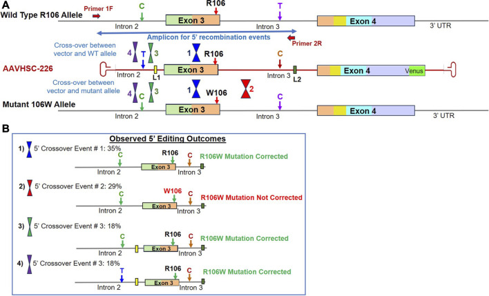
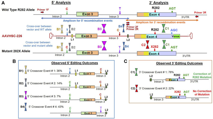
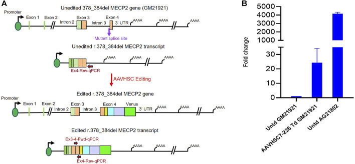
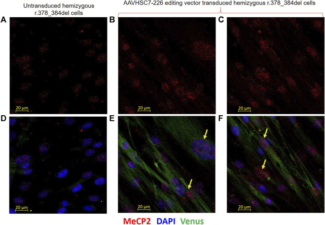
Rett syndrome is an acquired progressive neurodevelopmental disorder caused by de novo mutations in the X-linked MECP2 gene which encodes a pleiotropic protein that functions as a global transcriptional regulator and a chromatin modifier. Rett syndrome predominantly affects heterozygous females while affected male hemizygotes rarely survive. Gene therapy of Rett syndrome has proven challenging due to a requirement for stringent regulation of expression with either over- or under-expression being toxic. Ectopic expression of MECP2 in conjunction with regulatory miRNA target sequences has achieved some success, but the durability of this approach remains unknown. Here we evaluated a nuclease-free homologous recombination (HR)-based genome editing strategy to correct mutations in the MECP2 gene. The stem cell-derived AAVHSCs have previously been shown to mediate seamless and precise HR-based genome editing. We tested the ability of HR-based genome editing to correct pathogenic mutations in Exons 3 and 4 of the MECP2 gene and restore the wild type sequence while preserving all native genomic regulatory elements associated with MECP2 expression, thus potentially addressing a significant issue in gene therapy for Rett syndrome. Moreover, since the mutations are edited directly at the level of the genome, the corrections are expected to be durable with progeny cells inheriting the edited gene. The AAVHSC MECP2 editing vector was designed to be fully homologous to the target MECP2 region and to insert a promoterless Venus reporter at the end of Exon 4. Evaluation of AAVHSC editing in a panel of Rett cell lines bearing mutations in Exons 3 and 4 demonstrated successful correction and rescue of expression of the edited MECP2 gene. Sequence analysis of edited Rett cells revealed successful and accurate correction of mutations in both Exons 3 and 4 and permitted mapping of HR crossover events. Successful correction was observed only when the mutations were flanked at both the 5' and 3' ends by crossover events, but not when both crossovers occurred either exclusively upstream or downstream of the mutation. Importantly, we concluded that pathogenic mutations were successfully corrected in every Rett line analyzed, demonstrating the therapeutic potential of HR-based genome editing.

Keywords: MeCP2; Rett syndrome; adeno-associated virus; genome editing; homologous recombination.


Conflict of interest statement

SC is an advisor to and holds equity in Homology Medicines Inc. The remaining authors declare that the research was conducted in the absence of any commercial or financial relationships that could be construed as a potential conflict of interest.

Figures

FIGURE 1
FIGURE 1
Map and structure of MECP2 gene, protein and editing vector. (A) Genomic structure and protein domains of MECP2. MeCP2 protein domains and their corresponding locations in Exon 3 and 4 are color coded. (B) Map of the single-stranded AAVHSC-226 editing vector genome. The editing vector was designed to correct mutations in MECP2 Exons 3 and 4 and two unique linker sequences L1 and L2 were included in Introns 2 and 3, respectively. The editing vector was also designed to insert a promoterless T2A-Venus ORF cassette immediately downstream of Exon 4.
FIGURE 2
FIGURE 2
Specific Venus expression in edited R282X Rett syndrome fibroblasts. (A) Flow cytometric analysis of Venus expression in R282X fibroblasts 48 h post-transduction. Cells were transduced with AAVHSC15-226 at MOIs: 0, 150,000, 300,000 and 450,000. (B) Editing efficiency as a function of the MOI. Graph representing average editing efficiency in R282X cells at increasing vector dose (n = 3). Editing efficiency was calculated from specific Venus expression in transduced R282X fibroblasts.
FIGURE 3
FIGURE 3
Comparison of MECP2 editing by AAVHSC15 and AAVHSC7 in Rett syndrome cells. Heterozygous female R106W Rett syndrome fibroblasts (GM11273), hemizygous male S134C B-LCLs (GM17538) and heterozygous female R282X fibroblasts were transduced with either AAVHSC15-226 or AAVHSC7-226 editing vectors at MOI:150,000 (n = 3). Editing efficiency was determined by specific Venus expression 48 h post-transduction. Statistical analysis was performed using a two-sided, independent, two-sample t-test and the significance is shown (*p < 0.05; **p < 0.005).
FIGURE 4
FIGURE 4
Sequence analysis of the edited MECP2 gene in hemizygous male S134C Rett syndrome cells. (A) Editing of the MECP2 gene in S134C cells. Shown are the mutant S134C genome and the AAVHSC-226 editing vector. Also shown are: i) positions of SNPs which differed between the editing vector and the S134C genome, ii) location of linkers L1 and L2, iii) forward and reverse primers, iv) the S134C mutation. The primers used for TI analyses are depicted as thick red arrows. Primers 1F and 3R are specific for chromosomal sequences external to the homology arm. Primer 2R is specific for linker L2. Positions for HR crossover events between the mutant genome and the editing vector genome were identified by the presence or absence of markers in the edited genome and are depicted. (B) Observed 5′ editing outcomes. Two editing outcomes identified at the 5′ end based on the SNPs and linker sequences as markers are shown. (C) Observed 3′ editing outcomes. Two editing outcomes identified at the 3′ end based on the SNPs and linker sequences as markers are shown.
FIGURE 5
FIGURE 5
Sequence analysis of the edited MECP2 gene in heterozygous female R106W Rett syndrome cells. (A) Genome editing of the MECP2 gene in R106W cells. Shown are the WT R106 allele, AAVHSC-226 editing vector and the mutant 106W allele. Also shown are the locations of HR crossover events between the editing vector genome and either the WT R106 genome or the mutant 106W genome. The primers used for TI analyses are depicted as thick red arrows. Primer 1F is specific for chromosomal sequences external to the homology arms. Primer 2R is specific for linker L2. The crossover positions identified based on the presence or absence of markers in the edited genome and are depicted. (B) Observed editing outcomes. Four editing outcomes identified at the 5′ end based on the SNPs and linker sequences as markers are shown.
FIGURE 6
FIGURE 6
Sequence analysis of the edited MECP2 gene in heterozygous female R282X Rett syndrome cells. (A) Genome editing of the MECP2 gene in R282X cells. Shown are the WT R282 allele, AAVHSC-226 editing vector and the mutant 282X allele. Primers 1F and 3R are specific for chromosomal sequences external to the homology arms. Primers 2F and 2R are specific for linker L2. The crossover positions identified based on the presence or absence of markers in the edited genome and are depicted. (B) Observed 5′ editing outcomes. Four editing outcomes were identified at the 5′ end based on the presence or absence of specific SNPs and linkers as markers are shown. (C) Observed 3′ editing outcomes. Two editing outcomes identified at the 3′ end based on the SNPs and linker sequences as markers are shown.
FIGURE 7
FIGURE 7
Rescue of MECP2 expression in edited hemizygous GM21921 fibroblasts. (A) Schematic showing the r.378_384del mutation in the genome and transcript of GM21921 cells before and after genome editing. The cells contain a deletion at the Intron 3/Exon 4 splice junction resulting in formation of a new splice site and a premature termination codon. AAVHSC7-226 editing restored the wild type splice junction and Exon 4 sequence in r.378_384del cells. The primers used for qRT-PCR are depicted. The forward primer is complementary to the Exon 3/Exon 4 splice junction in the wild-type spliced transcript, which is deleted in GM12921 cells. The reverse primer anneals to Exon 4 downstream of the mutant premature termination codon. (B) Quantitation of restoration of MECP2 expression after editing. Shown is the fold-change in MECP2 transcript levels in AAVHSC7-226 edited GM21921 cells (AAVHSC7-226 Td GM21921) and wild-type AG21802 cells (Untd AG21802) compared with unedited GM21921 cells (Untd GM21921). The fold change represents the average of 2 experiments, with 3 replicates each. Bars represent the standard deviation.
FIGURE 8
FIGURE 8
Restoration of MeCP2 expression in edited male hemizygous r.378_384del fibroblasts. Intranuclear expression of MeCP2 is shown in male hemizygous fibroblasts after transduction with the AAVHSC7-226 editing vector. (A–C) MECP2 staining in untransduced and transduced r.378_384del fibroblasts. (A) Unedited cells show no MeCP2 expression due to a deletion in C-Ter. (B,C) MeCP2 expression is observed in nuclei of r.378_384del fibroblasts 48 h after transduction with the AAVHSC7-226 editing vector. (D) No Venus expression is observed in untransduced cells. (E, F) show Venus expression in the cytoplasm after overlay of staining with an anti-GFP antibody. Also shown in (D–F) is the overlay with DAPI staining. Yellow arrows: examples of cells expressing both MeCP2 and Venus.

Similar articles

Cited by

References

    1. Acampa M., Guideri F. (2006). Cardiac disease and Rett syndrome. Arch. Dis. Child. 91 (5), 440–443. 10.1136/adc.2005.090290 - DOI - PMC - PubMed
    1. Adikusuma F., Piltz S., Corbett M. A., Turvey M., McColl S. R., Helbig K. J., et al. (2018). Large deletions induced by Cas9 cleavage. Nature 560 (7717), E8–E9. 10.1038/s41586-018-0380-z - DOI - PubMed
    1. Agarwal N., Becker A., Jost K. L., Haase S., Thakur B. K., Brero A., et al. (2011). MeCP2 Rett mutations affect large scale chromatin organization. Hum. Mol. Genet. 20 (21), 4187–4195. 10.1093/hmg/ddr346 - DOI - PubMed
    1. Amir R. E., Van den Veyver I. B., Wan M., Tran C. Q., Francke U., Zoghbi H. Y. (1999). Rett syndrome is caused by mutations in X-linked MECP2, encoding methyl-CpG-binding protein 2. Nat. Genet. 23 (2), 185–188. 10.1038/13810 - DOI - PubMed
    1. Ausio J. (2016). MeCP2 and the enigmatic organization of brain chromatin. Implications for depression and cocaine addiction. Clin. Epigenetics 8, 58. 10.1186/s13148-016-0214-5 - DOI - PMC - PubMed

LinkOut - more resources