. 2024 Jan 12;3(3):421-430.
doi: 10.1039/d3sd00246b. eCollection 2024 Mar 14.

SARS-CoV-2 recombinase polymerase amplification assay with lateral flow readout and duplexed full process internal control

Affiliations

SARS-CoV-2 recombinase polymerase amplification assay with lateral flow readout and duplexed full process internal control

Coleman D Martin et al. Sens Diagn. .

Abstract

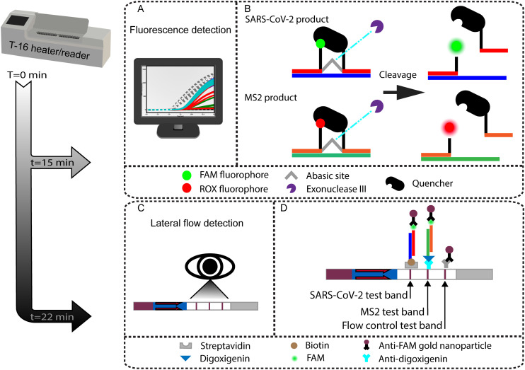
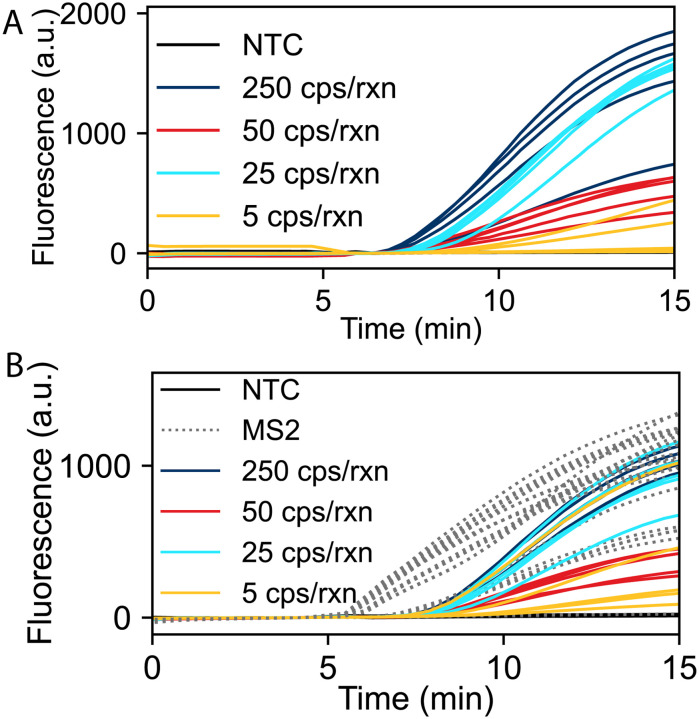
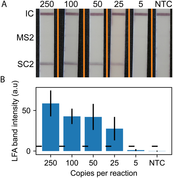
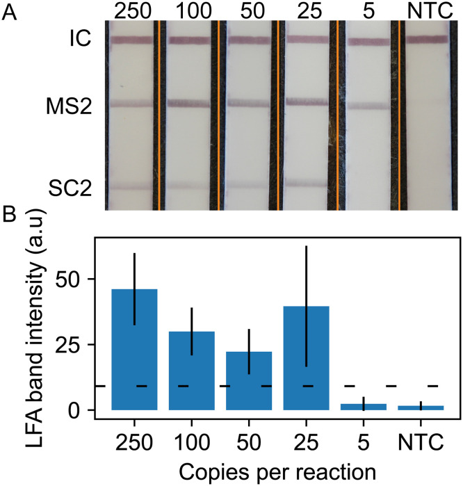
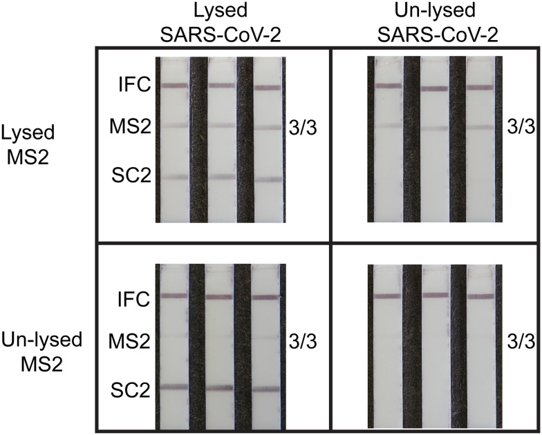
Nucleic acid amplification tests for the detection of SARS-CoV-2 have been an important testing mechanism for the COVID-19 pandemic. While these traditional nucleic acid diagnostic methods are highly sensitive and selective, they are not suited to home or clinic-based uses. Comparatively, rapid antigen tests are cost-effective and user friendly but lack in sensitivity and specificity. Here we report on the development of a one-pot, duplexed reverse transcriptase recombinase polymerase amplification SARS-CoV-2 assay with MS2 bacteriophage as a full process control. Detection is carried out with either real-time fluorescence or lateral flow readout with an analytical sensitivity of 50 copies per reaction. Unlike previously published assays, the RNA-based MS2 bacteriophage control reports on successful operation of lysis, reverse transcription, and amplification. This SARS-CoV-2 assay features highly sensitive detection, visual readout through an LFA strip, results in less than 25 minutes, minimal instrumentation, and a useful process internal control to rule out false negative test results.


Conflict of interest statement

There are no conflicts to declare.

Figures

Fig. 1
Fig. 1. Illustration of the duplexed RT-RPA assay with either real-time fluorescence or LFA-based detection. (A) The one-pot RT-RPA assay can be read out in real time using a fluorometer held at a constant 39 °C. (B) Mechanism of exonuclease cleaved fluorophore and quencher pairs for real-time multiplexed detection. When the SARS-CoV-2 amplicon is cleaved, the FAM fluorophore is detected. When the MS2 product is cleaved, the ROX fluorophore is detected. (C) Duplexed endpoint detection is also possible by LFA. (D) Design of the LFA. The LFA test strip is comprised of three ligand–receptor binding domains and includes the flow control line, labeled MS2 process control test line, and labeled SARS-CoV-2 test line.
Fig. 2
Fig. 2. RT-RPA amplification of SARS-CoV-2 RNA. The number of RNA target copies given per reaction. (A) Single target detection of SARS-CoV-2 (N = 5). (B) Duplexed detection of SARS-CoV-2 and MS2 (N = 5). All reactions containing MS2 RNA internal control amplified. Data shown with a baseline correction by subtracting the fluorescence value at 4 minutes.
Fig. 3
Fig. 3. Monoplexed LFA analysis. A) Representative lateral flow test strip readouts for RT-RPA assays with varying SARS-CoV-2 RNA input copy numbers. Orange lines denote composite image, strips taken from separate images and merged into one. B) Average (N = 5) SARS-CoV-2 test line intensity plotted with error bars representing one standard deviation (N = 5 for all concentrations). The horizontal dotted line is the positive test threshold defined as the NTC SARS-CoV-2 test line average intensity plus three standard deviations.
Fig. 4
Fig. 4. Duplexed LFA analysis. A) Representative test output. Orange lines denote composite image, strips taken from separate images and merged into one. B) Average (N = 5) SARS-CoV-2 test line intensity. Horizontal dotted line is the positive test threshold defined as the NTC SARS-CoV-2 test line average intensity plus three standard deviations.
Fig. 5
Fig. 5. Images of LFA strips demonstrating the successful lysis and RT-RPA of SARS-CoV-2 and the MS2 internal control. As expected, samples containing unlysed SARS-CoV-2 or MS2 are not successfully amplified and detected. The top row demonstrates true positive results (left) and true negative results (right) as the MS2 test line is detectable. The bottom left image demonstrates a failure of the MS2 internal control, even though SARS-CoV-2 is still detected. The bottom right image shows a complete assay failure where no banding is detected. Reading a valid test would require one of the two images from the top row. The bottom row demonstrates a failure in the internal control and an invalid test.

Similar articles

Cited by

References

    1. Phillips N. The Coronavirus Is Here to Stay — Here's What That Means. Nature. 2021;590(7846):382–384. doi: 10.1038/d41586-021-00396-2. doi: 10.1038/d41586-021-00396-2. - DOI - DOI - PubMed
    1. Peeling R. W. Heymann D. L. Teo Y.-Y. Garcia P. J. Diagnostics for COVID-19: Moving from Pandemic Response to Control. Lancet. 2022;399(10326):757–768. doi: 10.1016/S0140-6736(21)02346-1. doi: 10.1016/S0140-6736(21)02346-1. - DOI - DOI - PMC - PubMed
    1. Puhach O. Meyer B. Eckerle I. SARS-CoV-2 Viral Load and Shedding Kinetics. Nat. Rev. Microbiol. 2022;21:147–161. doi: 10.1038/s41579-022-00822-w. - DOI - PMC - PubMed
    1. Brihn A. Chang J. OYong K. Balter S. Terashita D. Rubin Z. Yeganeh N. Diagnostic Performance of an Antigen Test with RT-PCR for the Detection of SARS-CoV-2 in a Hospital Setting — Los Angeles County, California, June–August 2020. MMWR Morb. Mortal. Wkly. Rep. 2021;70(19):702–706. doi: 10.15585/mmwr.mm7019a3. doi: 10.15585/mmwr.mm7019a3. - DOI - DOI - PMC - PubMed
    1. Drain P. K. Rapid Diagnostic Testing for SARS-CoV-2. N. Engl. J. Med. 2022;386(3):264–272. doi: 10.1056/NEJMcp2117115. doi: 10.1056/NEJMcp2117115. - DOI - DOI - PMC - PubMed

LinkOut - more resources