. 2024 Mar 1:15:1287111.
doi: 10.3389/fgene.2024.1287111. eCollection 2024.

Genome-wide identification of dysregulated alternative splicing and RNA-binding proteins involved in atopic dermatitis

Affiliations

Genome-wide identification of dysregulated alternative splicing and RNA-binding proteins involved in atopic dermatitis

Yaqi Yang et al. Front Genet. .

Abstract

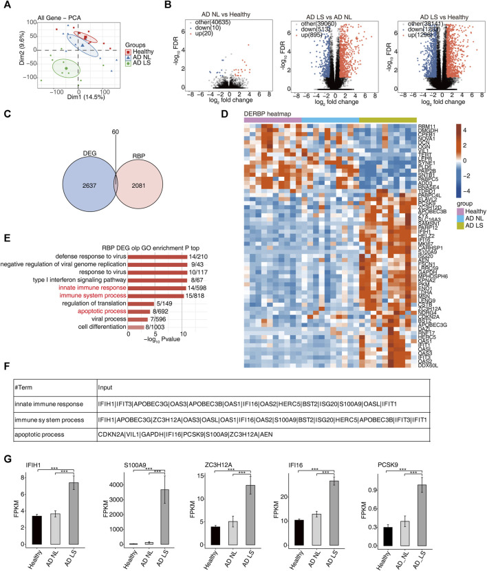
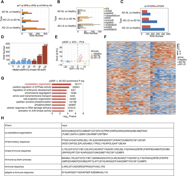
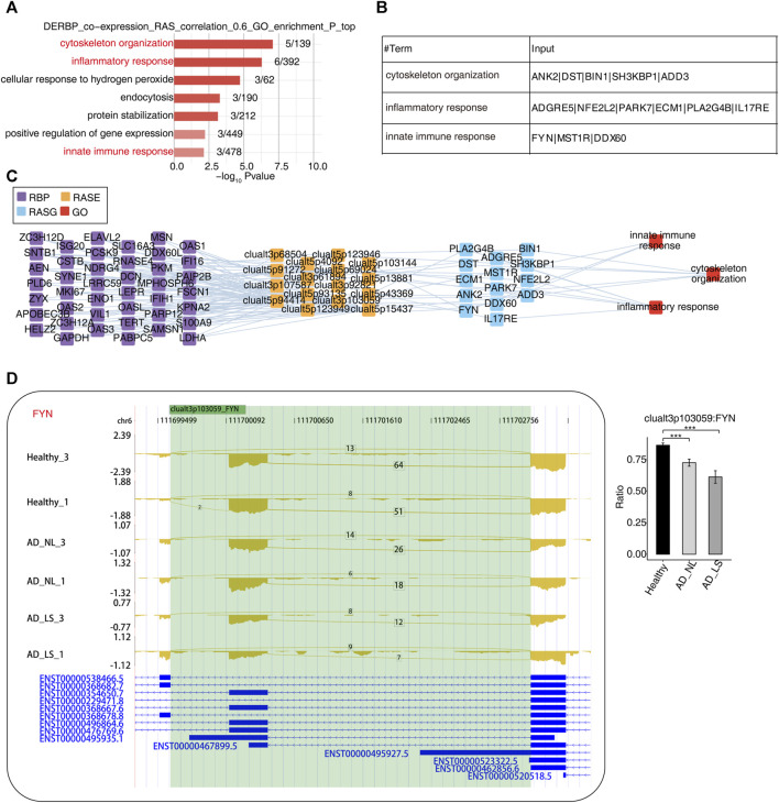
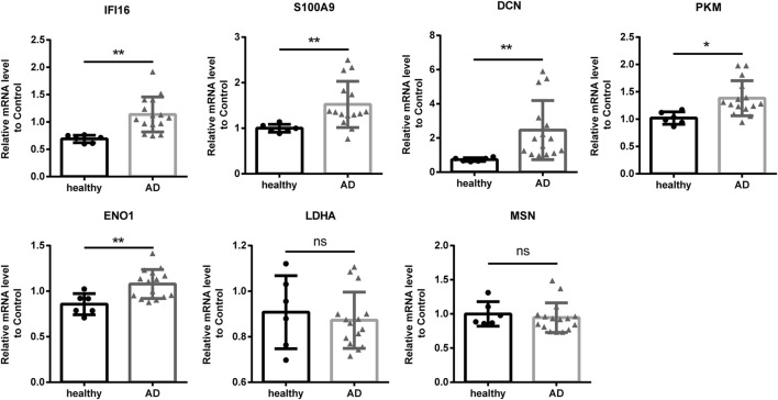
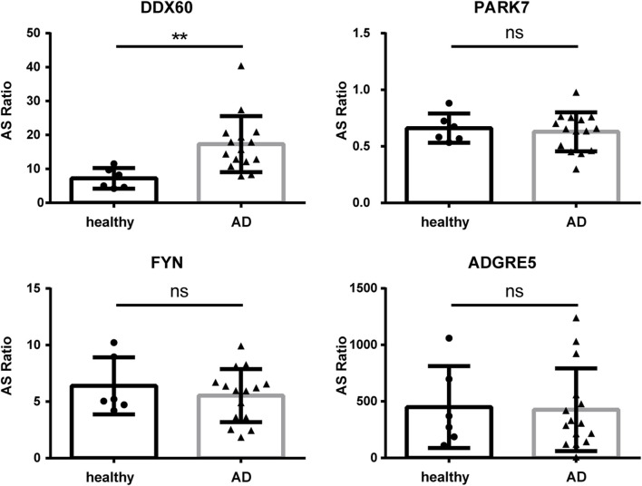
Objectives: We explored the role and molecular mechanisms of RNA-binding proteins (RBPs) and their regulated alternative splicing events (RASEs) in the pathogenesis of atopic dermatitis (AD). Methods: We downloaded RNA-seq data (GSE121212) from 10 healthy control skin samples (healthy, Ctrl), 10 non-lesional skin samples with AD damage (non-lesional, NL), and 10 lesional skin samples with AD damage (lesional, LS). We performed the analysis of differentially expressed genes (DEGs), differentially expressed RBPs (DE-RBPs), alternative splicing (AS), functional enrichment, the co-expression of RBPs and RASEs, and quantitative polymerase chain reaction (qPCR). Results: We identified 60 DE-RBP genes by intersecting 2141 RBP genes from existing reports with overall 2697 DEGs. Most of the DE-RBP genes were found to be upregulated in the AD LS group and related to immune and apoptosis pathways. We observed different ASEs and RASEs among the healthy, AD NL, and AD LS groups. In particular, alt3p and alt5p were the main ASEs and RASEs in AD NL and AD LS groups, compared to the healthy group. Furthermore, we constructed co-expression networks of DE-RBPs and RAS, with particular enrichment in biological pathways including cytoskeleton organization, inflammation, and immunity. Subsequently, we selected seven genes that are commonly present in these three pathways to assess their expression levels in the peripheral blood mononuclear cells (PBMCs) from both healthy individuals and AD patients. The results demonstrated the upregulation of four genes (IFI16, S100A9, PKM, and ENO1) in the PBMCs of AD patients, which is highly consistent with DE-RBP genes analysis. Finally, we selected four RAS genes regulated by RBPs that were related to immune pathways and examined their RASEs in PBMCs from both AD patients and healthy controls. The results revealed an increased percentage of RASEs in the DDX60 gene in AD, which is highly consistent with AS analysis. Conclusion: Dysregulated RBPs and their associated RASEs may have a significant regulatory role in the development of AD and could be potential therapeutic targets in the future.

Keywords: RNA-binding proteins; alternative splicing; atopic dermatitis; genome-wide analysis; immune and inflammatory response.


Conflict of interest statement

The authors declare that the research was conducted in the absence of any commercial or financial relationships that could be construed as a potential conflict of interest.

Figures

FIGURE 1
FIGURE 1
Transcriptome analysis of DE-RBPs in atopic dermatitis lesional and atopic dermatitis non-lesional samples compared with healthy samples. (A) PCA based on the FPKM value of all detected genes. The ellipse for each group is the confidence ellipse. (B) Volcano plots presenting all DEGs in the AD_LS vs. healthy, AD_NL vs. healthy, and AD_LS vs. AD_NL comparison groups. FDR ≤0.05 and FC ≥ 2 or ≤0.5. (C) Venn diagram showing the overlap of DEGs and RBPs. (D) Heatmap diagram showing the expression profile of specific DE-RBPs in the three comparison groups. (E) Bar plot showing the most enriched GO biological process results of specific DE-RBPs in the three comparison groups. The color scale shows the row-scaled significance (-log10-corrected p-value) of the terms. (F) Sheet showing the genes enriched in the key GO pathway. (G) Bar plot showing the expression pattern and statistical difference of DE-RBPs. Error bars represent mean ± SEM. ***p-value <0.001.
FIGURE 2
FIGURE 2
A large number of alternative splicing events related to inflammatory and immune responses were identified in atopic dermatitis development. (A) Bar plot showing several regulatory RAS detected using the SUVA in each group. (B) Splice junction constituting RAS events detected using the SUVA was annotated to classical AS event types. The number of each classical AS event type is shown with a bar plot. (C) Complex and simple variable splicing event detection cases. (D) Bar plot showing RAS with different pSAR values. RAS whose pSAR (Reads proportion of SUVA AS event) ≥ 50% were labeled. (E) PCA based on RAS with pSAR ≥50%. The ellipse for each group is the confidence ellipse. (F) Heatmap showing the splicing ratio of specific RAS (pSAR ≥50%) in the three groups. (G) Bar plot exhibiting the most enriched GO biological process results of specific RAS (pSAR ≥50%) genes in the three groups. The color scale shows the row-scaled significance (-log10 corrected p-value) of the terms. (H) Sheet exhibiting several key GO biological process results of RAS (pSAR ≥50%).
FIGURE 3
FIGURE 3
Construction of a co-dysregulated network between RNA-binding proteins and unstable plaque-specific RAS in atopic dermatitis development. (A) Bar plot showing the top 10 most enriched GO biological process results of specific DE-RBPs co-expressed by specific RAS. The color scale shows the row-scaled significance (-log10 corrected p-value) of the terms. (B) Sheet exhibiting several key GO biological process results of specific DE-RBPs co-expressed by specific RAS. (C) Co-expression analysis of specific DE-RBPs and specific RAS of key GO biological process results. Cutoffs of p-value ≤0.01 and Pearson coefficient ≥0.6 or ≤ −0.6 were applied to identify the co-expression pairs. The network shows the co-expressed GO pathway for specific DE-RBPs (the far left part) and specific RAS (the middle left part). The top enriched GO terms of RASGs (the middle right part) are shown in red (the far right part). (D) Reads distribution diagram showing clualt3p103059 FYN. Bar plot showing the splicing ratio of clualt3p103059 FYN on the right. Error bars represent mean ± SEM. *: p-value ≤0.05, **: p-value ≤0.01, and ***: p-value ≤0.001.
FIGURE 4
FIGURE 4
Validation of important RBPs in PBMCs in clinical samples. Results of the qPCR of the gene expression of seven RPBs. RBP, RNA-binding protein; qPCR, quantitative polymerase chain reaction. *: p-value ≤0.05; **: p-value ≤0.01.
FIGURE 5
FIGURE 5
Validation of important RAS genes in PBMCs in clinical samples. qPCR was performed to detect the splicing ratio of four RAS genes. RAS, regulatory alternative splicing; qPCR, quantitative polymerase chain reaction. **: p-value ≤0.01.

Similar articles

References

    1. Akdis C. A., Akdis M., Trautmann A., Blaser K. (2000). Immune regulation in atopic dermatitis. Curr. Opin. Immunol. 12 (6), 641–646. 10.1016/s0952-7915(00)00156-4 - DOI - PubMed
    1. Akira S., Maeda K. (2021). Control of RNA stability in immunity. Annu. Rev. Immunol. 39, 481–509. 10.1146/annurev-immunol-101819-075147 - DOI - PubMed
    1. Ali F., Vyas J., Finlay A. Y. (2020). Counting the burden: atopic dermatitis and health-related quality of life. Acta Derm. Venereol. 100 (12), adv00161. 10.2340/00015555-3511 - DOI - PMC - PubMed
    1. Angiolilli C., Leijten E., Bekker C., Eeftink E., Giovannone B., Nordkamp M. O., et al. (2022). ZFP36 family members regulate the proinflammatory features of psoriatic dermal fibroblasts. J. Invest. Dermatol 142 (2), 402–413. 10.1016/j.jid.2021.06.030 - DOI - PubMed
    1. Astolfi A., Cipriani F., Messelodi D., De Luca M., Indio V., Di Chiara C., et al. (2021). Filaggrin loss-of-function mutations are risk factors for severe food allergy in children with atopic dermatitis. J. Clin. Med. 10 (2), 233. 10.3390/jcm10020233 - DOI - PMC - PubMed