. 2024 Feb 16;27(4):109251.
doi: 10.1016/j.isci.2024.109251. eCollection 2024 Apr 19.

Transcriptome regulation by PARP13 in basal and antiviral states in human cells

Affiliations

Transcriptome regulation by PARP13 in basal and antiviral states in human cells

Veronica F Busa et al. iScience. .

Abstract

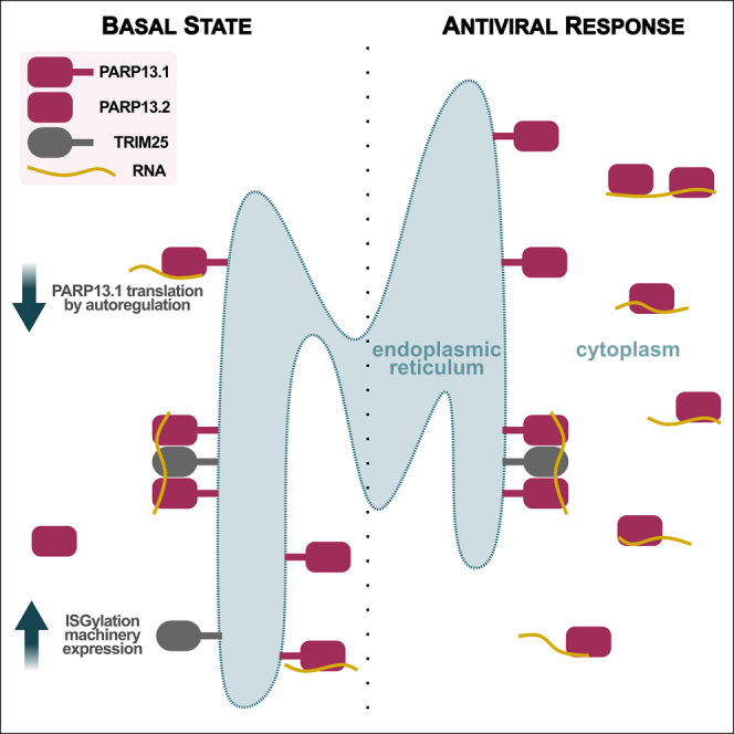
The RNA-binding protein PARP13 is a primary factor in the innate antiviral response, which suppresses translation and drives decay of bound viral and host RNA. PARP13 interacts with many proteins encoded by interferon-stimulated genes (ISG) to activate antiviral pathways including co-translational addition of ISG15, or ISGylation. We performed enhanced crosslinking immunoprecipitation (eCLIP) and RNA-seq in human cells to investigate PARP13's role in transcriptome regulation for both basal and antiviral states. We find that the antiviral response shifts PARP13 target localization, but not its binding preferences, and that PARP13 supports the expression of ISGylation-related genes, including PARP13's cofactor, TRIM25. PARP13 associates with TRIM25 via RNA-protein interactions, and we elucidate a transcriptome-wide periodicity of PARP13 binding around TRIM25. Taken together, our study implicates PARP13 in creating and maintaining a cellular environment poised for an antiviral response through limiting PARP13 translation, regulating access to distinct mRNA pools, and elevating ISGylation machinery expression.

Keywords: Cell biology; Immune response; Molecular biology.


Conflict of interest statement

G.W.Y. is co-founder, member of the Board of Directors, on the SAB, equity holder, and paid consultant for Locanabio and Eclipse BioInnovations. G.W.Y. is a visiting professor at the National University of Singapore. G.W.Y.’s interest(s) have been reviewed and approved by the University of California San Diego in accordance with its conflict-of-interest policies. The authors declare no other competing interests.

Figures

None
Graphical abstract
Figure 1
Figure 1
PARP13 expression affects expression of many constitutive transcripts and is required for transcriptomic upregulation of innate immune response (A) Log2 fold change expression data 24 h after treatment with +3p-RNA vs. +ssRNA in HEK293T WT and PARP13 KO cells (n = 2). A selection of innate immune response genes is highlighted in red. (B) Log2 fold change expression data from HEK293T WT vs. PARP13 KO cell 24 h after treatment with +3p-RNA or +ssRNA (n = 2). (C) Scaled average expression relative to WT + ssRNA measured by qPCR in WT HEK293T and PARP13 KO cells and normalized to GAPDH or TBP expression (n ≥ 3). Complete replicate information Figure S1B. (D) IC pattern weights across RNA-seq samples. The color scale represents values of the linear mixing matrix centered at 0 where columns of S contain the independent components, A is a linear mixing matrix, and data matrix X is considered to be a linear combination of non-Gaussian components such that X = SA. (E) Top GO terms that are enriched among genes that contribute to IC2, shown in (D). (F) Top GO terms that are enriched among genes that contribute to IC3, shown in (D).
Figure 2
Figure 2
PARP13 selectively binds PARP13.1 and regulates its translation via the 3′ UTR (A) Proportions of PARP13 binding sites that fall within the indicated gene regions of mRNAs. (B) Log2 fold-change RNA expression of PARP13-bound genes in PARP13 KO versus WT +ssRNA-treated cells, separated by what region of the transcript PARP13 binds. Transcripts where PARP13 binds multiple types of gene regions were excluded. Colors correspond to the gene region legend on the left. T-test, ∗∗ = p < 0.01. (C) RNA immunoprecipitation using GFP-trap beads against exogenous GFP-PARP13.1 in HEK293T cells. Error bars are +/− 1sd. (D) Aligned RNA-seq (top) and eCLIP-seq (middle) reads within PARP13 gene region for +ssRNA and +3p-RNA treatments with gray boxes surrounding the PARP13.1-specific (left) and PARP13.2-specific (right) 3′UTRs. Red and blue alignments correspond to duplicate experiments. Two PARP13 binding peaks located within the PARP13.1 3′UTR identified by CLIPper (Methods) are indicated below the aligned reads. Bottom: a splicing map of the two PARP13 isoforms, which are transcribed right to left. (E) Western blot of PARP13 expression in WT and PARP13 KO HEK293T cell lysates 24 h after +ssRNA or +3p-RNA treatment, with beta-actin as a loading control. Also Figure S2A. (F) Luciferase construct map and design of luciferase expression experiments. (G) Transcript levels of Renilla luciferase with either PARP13.1 or PARP13.2 3′UTR treated with ssRNA or 3p-RNA (n = 5). Within each experiment, Renilla expression was normalized to firefly expression and compared to normalized Renilla expression for a construct without a 3′UTR insert. ns = not significant. (H) Luminescence of Renilla luciferase (n = 3) with the same normalization as (G). Paired t-test, ∗∗ = p < 0.01; ns = not significant.
Figure 3
Figure 3
Antiviral response shifts PARP13 target localization but not binding preferences (A) Venn diagram of transcripts that have at least one significantly bound region across both treatments. (B) Dinucleotide enrichment within PARP13-bound regions, normalized to dinucleotide expression of all transcribed RNAs. (C) A hexameric motif calculated by HOMER that is enriched in samples with either +ssRNA (top) and +3p-RNA (bottom) treatments. (D) Proportions of previously identified cellular localizations for PARP13-bound transcripts across both treatments.
Figure 4
Figure 4
Genes associated with ISGylation are dysregulated in PARP13 KO cells (A) A schematic of the ISGylation pathway that includes ISG15, a ubiquitin-like protein; UBA7, the E1; UBE2L6, the E2; HERC5 and TRIM25, E3 ligases; and USP18 and USP41, which remove ISG15 from conjugated proteins. RIG-I, which is ubiquitinated by TRIM25, activates expression of ISG15. Genes that have been shown to promote PARP13 antiviral activity are highlighted in purple. Genes identified by the RNA-seq data to be down-regulated in PARP13 KO cells in control condition and upregulated upon mock viral infection in WT cells are highlighted in gold. Proteins shown to bind PARP13 are connected by dotted lines. (B) qPCR of ISGylation genes in untreated WT and PARP13 KO HEK293T cells (n = 3). UBA7 expression was too low and beyond the limit of detection. Error bars are +/− 1sd. (C) Western blot of PARP13 and TRIM25 expression in WT and PARP13 KO HEK293T cell lysates, with beta-actin as a loading control. Also Figure S2B. (D) PARP13 KD time course in WT HEK293T cells assessing the expression of ISGylation genes shown in (B) to have reduced expression in PARP13 KO cells. Error bars are +/− 1sd.
Figure 5
Figure 5
TRIM25 and PARP13 interact via both RNA-dependent and RNA-independent contacts (A) Venn diagram of previously identified TRIM25 targets from HeLa cells transiently expressing T7-tagged TRIM25 and all PARP13 targets from HEK293T cells. (B) Proportions of PARP13-bound transcripts across both treatments that are also targets of TRIM25 . (C) Spatial correlation of PARP13- and TRIM25-bound regions across the HEK293T transcriptome calculated by nearBynding. Black line indicates mean shuffled background signal with standard error (n = 10,000) and blue line represents mean spatial correlation signal with standard error (n = 3). Arrows are the same on the left and right plots and represent periodic regions of significant spatial correlation between TRIM25 and PARP13. Left: Correlation of TRIM25 binding peaks from HeLa cells and PARP13 binding peaks from ssRNA-treated HEK293T cells. Right: Correlation of TRIM25 binding peaks and PARP13 binding peaks from 3p-RNA-treated cells. (D) Three possible models of TRIM25 and PARP13 binding proximity to explain the periodic signal observed in the spatial correlation in (C). 1. Multiple TRIM25 proteins bind near, upstream, and downstream a single PARP13 protein. 2. Multiple PARP13 proteins bind near, upstream, and downstream of a single TRIM25 protein. Or 3. a combination of 1 and 2, where multiple binding events of both proteins are proximal. (E) Auto-correlation of TRIM25 binding. Spatial correlation of a binding site to itself equals 1 at position 0. The same auto-correlation with a magnified y-axis is shown in the top right. (F and G) Auto-correlation of PARP13 binding with ssRNA treatment (F) and 3p-RNA treatment (G). Spatial correlation of a binding site to itself equals 1 at position 0, but peaks upstream and downstream show additional PARP13 binding sites nearby. The same auto-correlation with a magnified y-axis is shown in the top right. (H) An example PARP13 and TRIM25 target, ATP6AP1. A splicing map of ATP6AP1 is highlighted by a red box corresponding to the region for which aligned PARP13 eCLIP-seq reads are shown for two replicates in ssRNA-treated cells. Significant binding peaks are indicated below for PARP13 (blue) and TRIM25 (red). (I) Western blot for GFP and TRIM25 expression from co-immunoprecipitation using GFP-trap beads against exogenous GFP-PARP13 constructs in HEK293T cells. Also Figure S2C.

Similar articles

Cited by

References

    1. Ficarelli M., Neil S.J.D., Swanson C.M. Targeted Restriction of Viral Gene Expression and Replication by the ZAP Antiviral System. Annu. Rev. Virol. 2021;8:265–283. doi: 10.1146/annurev-virology-091919-104213. - DOI - PubMed
    1. Hayakawa S., Shiratori S., Yamato H., Kameyama T., Kitatsuji C., Kashigi F., Goto S., Kameoka S., Fujikura D., Yamada T., et al. ZAPS is a potent stimulator of signaling mediated by the RNA helicase RIG-I during antiviral responses. Nat. Immunol. 2011;12:37–44. doi: 10.1038/ni.1963. - DOI - PubMed
    1. Chen S., Xu Y., Zhang K., Wang X., Sun J., Gao G., Liu Y. Structure of N-terminal domain of ZAP indicates how a zinc-finger protein recognizes complex RNA. Nat. Struct. Mol. Biol. 2012;19:430–435. doi: 10.1038/nsmb.2243. - DOI - PubMed
    1. Schwerk J., Soveg F.W., Ryan A.P., Thomas K.R., Hatfield L.D., Ozarkar S., Forero A., Kell A.M., Roby J.A., So L., et al. RNA-binding protein isoforms ZAP-S and ZAP-L have distinct antiviral and immune resolution functions. Nat. Immunol. 2019;20:1610–1620. doi: 10.1038/s41590-019-0527-6. - DOI - PMC - PubMed
    1. Zhu Y., Wang X., Goff S.P., Gao G. Translational repression precedes and is required for ZAP-mediated mRNA decay. EMBO J. 2012;31:4236–4246. doi: 10.1038/emboj.2012.271. - DOI - PMC - PubMed