. 2024 Mar 1:15:1346687.
doi: 10.3389/fimmu.2024.1346687. eCollection 2024.

Identification of immunological patterns characterizing immune-related psoriasis reactions in oncological patients in therapy with anti-PD-1 checkpoint inhibitors

Affiliations

Identification of immunological patterns characterizing immune-related psoriasis reactions in oncological patients in therapy with anti-PD-1 checkpoint inhibitors

Martina Morelli et al. Front Immunol. .

Abstract

Introduction: Immunotherapy with biologics targeting programmed cell death protein-1 (PD-1) is highly effective in the treatment of various malignancies. Nevertheless, it is frequently responsible for unexpected cutaneous manifestations, including psoriasis-like dermatitis. The pathogenesis of anti-PD-1-induced psoriasis has yet to be clarified, even though it is plausible that some innate and adaptive immunity processes are in common with canonical psoriasis. The genetic predisposition to psoriasis of patients could also be a contributing factor. Here, we investigated the immunological and genetic profiles of two patients with metastatic melanoma and one patient affected by lung cancer, who developed severe psoriasis after receiving anti-PD-1 nivolumab therapy.

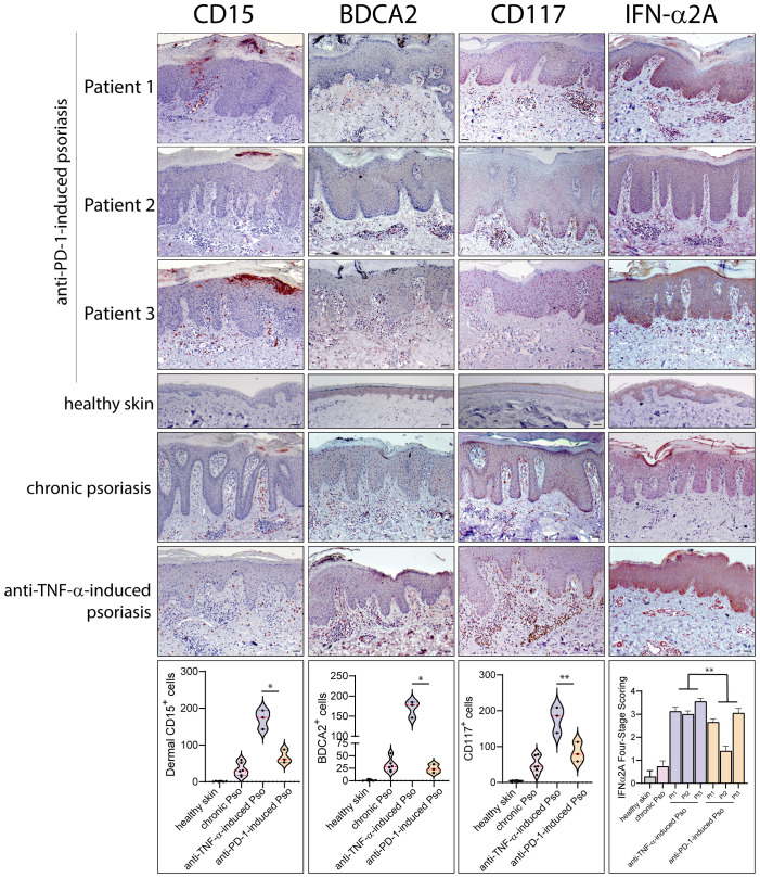
Methods: The immune patterns of the three patients were compared with those detectable in classical, chronic plaque-type psoriasis or paradoxical psoriasis induced by anti-TNF-α therapy, mostly sustained by adaptive and innate immunity processes, respectively. Therefore, immunohistochemistry and mRNA analyses of innate and adaptive immunity molecules were conducted on skin biopsy of patients. Genetic analysis of polymorphisms predisposing to psoriasis was carried out by NGS technology.

Results: We found that anti-PD-1-induced psoriasis showed immunological features similar to chronic psoriasis, characterized by the presence of cellular players of adaptive immunity, with abundant CD3+, CD8+ T cells and CD11c+ dendritic cells infiltrating skin lesions, and producing IL-23, IL-6, TNF-α, IFN-γ and IL-17. On the contrary, a lower number of innate immunity cells (BDCA2+ plasmacytoid dendritic cells, CD15+ neutrophils, CD117+ mast cells) and reduced IFN-α/β, lymphotoxin (LT)-α/β, were observed in anti-PD-1-induced psoriasis lesions, as compared with anti-TNF-α-induced paradoxical psoriasis. Importantly, the disintegrin and metalloprotease domain containing thrombospondin type 1 motif-like 5 (ADAMTSL5) psoriasis autoantigen was significantly upregulated in psoriasis lesions of anti-PD-1-treated patients, at levels comparable with chronic plaque-type psoriasis. Finally, NGS analysis revealed that all patients carried several allelic variants in psoriasis susceptibility genes, such as HLA-C, ERAP1 and other genes of the major psoriasis susceptibility PSORS1 locus.

Discussion: Our study showed that adaptive immunity predominates over innate immunity in anti-PD-1-induced psoriasis lesions, consistently with the local ADAMTSL5 overexpression. The presence of numerous SNPs in psoriasis susceptibility genes of the three patients also suggested their strong predisposition to the disease.

Keywords: adaptive immunity; anti-PD-1 therapy; immune-related cutaneous adverse event (ircAE); innate immunity; melanoma; paradoxical skin reactions; psoriasis.


Conflict of interest statement

The authors declare that the research was conducted in the absence of any commercial or financial relationships that could be construed as a potential conflict of interest.

Figures

Figure 1
Figure 1
Clinical and histological presentation of psoriasis induced by anti-PD-1 therapy. Cutaneous manifestations of patients 1 and 2 affected by lung cancer and melanoma, respectively, presenting psoriasis reaction after receiving anti-PD-1 treatment. (A) Patient 1, panels i-iii show paradoxical erythemato-squamous plaques localized on the trunk, arms and lower limbs. Patient 1 also developed generalized bullous pemphigoid, arising after treatment with nivolumab (panels iv-v). (B) Patient 2 shows squamous plaques on the elbows and upper limbs (i) and on the trunk (ii), concomitantly with vitiligo (ii and iii). (C) H&E staining for the corresponding histology specimens of patients 1 (i), patient 2 (ii) and patient 3 (iii) was also performed. Scale bars, 20 μm.
Figure 2
Figure 2
Paradoxical psoriasis induced by PD-1 blockade show a prominent infiltrate of CD3+, CD8+ T cells, CD11c+ DC and IL-17+ cells, at levels and patterns of distribution similar to chronic psoriasis. Leukocyte subpopulations were characterized by immunohistochemistry in paradoxical psoriasis lesions induced by anti-PD-1 (Patient 1, 2 and 3), and compared to those present in chronic plaque-type psoriasis (n=6 patients) and in paradoxical psoriasis induced by anti-TNF-α therapy (n=3). The distribution of numerical data relative to cell immunoreactivity for CD3 (red staining), CD8 (red), CD11C (brown), and IL-17A (red staining) in the three types of psoriasis reactions, are represented in the violin plots. Immunohistochemistry analysis of anti-PD-1 psoriasis skin reactions obtained from patient 1, patient 2 and 3 shows similar numbers of CD3+ cells and higher number of epidermal CD8+, dermal CD11C+, and IL-17A+ cells, when compared with paradoxical psoriasis induced by anti-TNF-α. Chronic psoriasis and anti-PD-1-induced psoriasis showed similar values in immunoreactive cells. No immunoreactivities were observed in skin samples from healthy donors (n=6). Arrows indicate CD8+ T cells localized within epidermis. Slides were analyzed by two pathologists with experience in dermatology. Positive cells were counted in five adjacent fields at a total magnification of ×200. For chronic or anti-TNF-α-induced psoriasis, one representative set of staining is shown. For each patient, one out of three representative stainings is shown. *p < 0.05, **p < 0.01 versus anti-TNF-α-induced psoriasis. Scale bars, 40 μm.
Figure 3
Figure 3
Innate immunity patterns are lacking in anti-PD-1-induced psoriasis. Innate immunity cell subpopulations were characterized by immunohistochemistry in paradoxical psoriasis lesions induced by anti-PD-1 (Patient 1, 2 and 3), and compared to those present in chronic plaque-type psoriasis (n=6 patients) and in paradoxical psoriasis induced by anti-TNF-α therapy (n=3). The distribution of numerical data relative to cell immunoreactivity for CD15 (red staining), BDCA2 (brown) and c-kit/CD117 (red staining) in the three types of psoriasis reactions are represented in the violin plots. Graph shows the mean ± SD of semiquantitative, four-stage scoring, ranging from negative immunoreactivity (0) to strong immunoreactivity (4+) of IFN-α2A staining (red). The infiltrate of dermal CD15+ neutrophils BDCA2+ pDC, and c-kit/CD117+ mast cells, was less abundant in immune-related psoriasis induced by anti-PD-1 as compared to paradoxical psoriasis induced by anti-TNF-α. IFN-α2A also was less abundant in psoriasiform reactions of anti-PD-1-treated patients, as compared to paradoxical psoriasis induced by anti-TNF-α. Similar immunoreactivity values were observed in anti-PD-1-induced and chronic psoriasis. No significant immunoreactivities were observed in skin samples from healthy donors (n=6). Slides were analyzed by two pathologists with experience in dermatology. Positive cells were counted in five adjacent fields at a total magnification of ×200. For chronic or anti-TNF-α-induced psoriasis, one representative set of reactions is shown. For each patient, one out of three representative staining is shown. *p < 0.05, **p < 0.01 versus anti-TNF-α-induced psoriasis. Scale bars, 40 μm.
Figure 4
Figure 4
Inflammatory cytokines typical of chronic psoriasis are overexpressed in immune-related psoriasis reactions to anti-PD-1. (A) mRNA expression of IL-17A, IL-22, IFN-γ, TNF-α, IL-6 and IL-23 was analyzed by real-time PCR in psoriasis lesions induced by anti-PD-1 (Pt 1, Pt2 and Pt3), and compared to those present in skin biopsies from lesional (Pso LS) and non-lesional (Pso NLS) skin of psoriatic patients (n=6) and in paradoxical psoriasis induced by anti-TNF-α (n=3). Healthy skin from healthy donors (Healthy, n=6) was also analyzed. Levels of all mRNAs were significantly higher in the skin of the three patients, as compared to psoriasis-like reactions to anti-TNF-α, and similar to those detected in chronic psoriasis plaques. (B) Real-time PCR analysis of selected innate immunity molecules showed that IFN-β, LT-α and LT-β mRNAs are less expressed in psoriasis lesions induced by anti-PD-1, as compared to psoriasis reactions to anti-TNF-α. mRNA values were normalized to β2M mRNA. All data shown are the mean of three different experiments ± SD. Statistical significance was assessed by paired Student’s t test, *p ≤ 0.05, **p ≤ 0.01.
Figure 5
Figure 5
ADAMTSL5 psoriasis autoantigen is overexpressed in psoriasis lesions induced by nivolumab treatment, as well as in melanoma tissues. (A) Immunohistochemistry for ADAMTSL5 was conducted on psoriasis lesions induced by anti-PD-1 (Patient 1, 2 and 3), and compared to those present in chronic plaque-type psoriasis (n=6 patients) and in psoriasis induced by anti-TNF-α therapy (n=3). ADAMTSL5 immunoreactivity was mainly present in paradoxical psoriasis to anti-PD-1, in keratinocytes and in scattered basal cells with the morphology of melanocytes. It was also highly expressed in most dermal infiltrating cells, localized in the papillary and mid dermis, as well in perivascular and endothelial cells. Panels iv, v and vi represent the insets of panels i, ii, and iii, respectively, at a magnification of x200. ADAMTSL5 expression pattern observed in psoriasis to anti-PD-1 was similar to that observed in plaque-type psoriasis (vii). Anti-TNF-α -induced psoriasis lesions were negative (viii). (B) For patient 2, ADAMTSL5 was also detected in non-lesional (3-cm distant area from tumor lesion) (i), perilesional (ii) and lesional (iii-v) skin specimens obtained from the primary melanoma and melanoma skin metastasis (vi). Numerous ADAMTSL5+ melanophages/macrophages infiltrated melanoma tissues (arrows, iii-vi). (C) ADAMTSL5 expression was absent in both perilesional (i) and lesional (ii) skin of anti-PD-1-induced vitiligo developed by patient 2 after nivolumab treatment. For each specimen, one out of three representative staining is shown. Scale bars, 40 μm.

References

    1. Robert C, Long GV, Brady B, Dutriaux C, Maio M, Mortier L, et al. . Nivolumab in previously untreated melanoma without BRAF mutation. N Engl J Med. (2015) 372:320–30. doi: 10.1056/NEJMoa1412082 - DOI - PubMed
    1. Hellmann MD, Paz-Ares L, Bernabe Caro R, Zurawski B, Kim SW, Carcereny Costa E, et al. . Nivolumab plus ipilimumab in advanced non-small-cell lung cancer. N Engl J Med. (2019) 381:2020–31. doi: 10.1056/NEJMoa1910231 - DOI - PubMed
    1. Xing P, Zhang F, Wang G, Xu Y, Li C, Wang S, et al. . Incidence rates of immune-related adverse events and their correlation with response in advanced solid tumours treated with NIVO or NIVO+IPI: a systematic review and meta-analysis. J Immunother Cancer. (2019) 7:341–50. doi: 10.1186/s40425-019-0779-6 - DOI - PMC - PubMed
    1. Shah NJ, Lacouture ME. Dermatologic immune-related adverse events to checkpoint inhibitors in cancer. J Allergy Clin Immunol. (2023) 151:407–09. doi: 10.1016/j.jaci.2022.11.015 - DOI - PubMed
    1. Maillard A, Pastor D, Merat R. Anti-PD-1-induced hidradenitis suppurativa. Dermatopathology. (2021) 8:37–9. doi: 10.3390/dermatopathology8010007 - DOI - PMC - PubMed

Publication types

MeSH terms