. 2024 Mar 10:27:39-47.
doi: 10.1016/j.reth.2024.02.005. eCollection 2024 Dec.

A passage-free, simplified, and scalable novel method for iPSC generation in three-dimensional culture

Affiliations

A passage-free, simplified, and scalable novel method for iPSC generation in three-dimensional culture

Masaya Tsukamoto et al. Regen Ther. .

Abstract

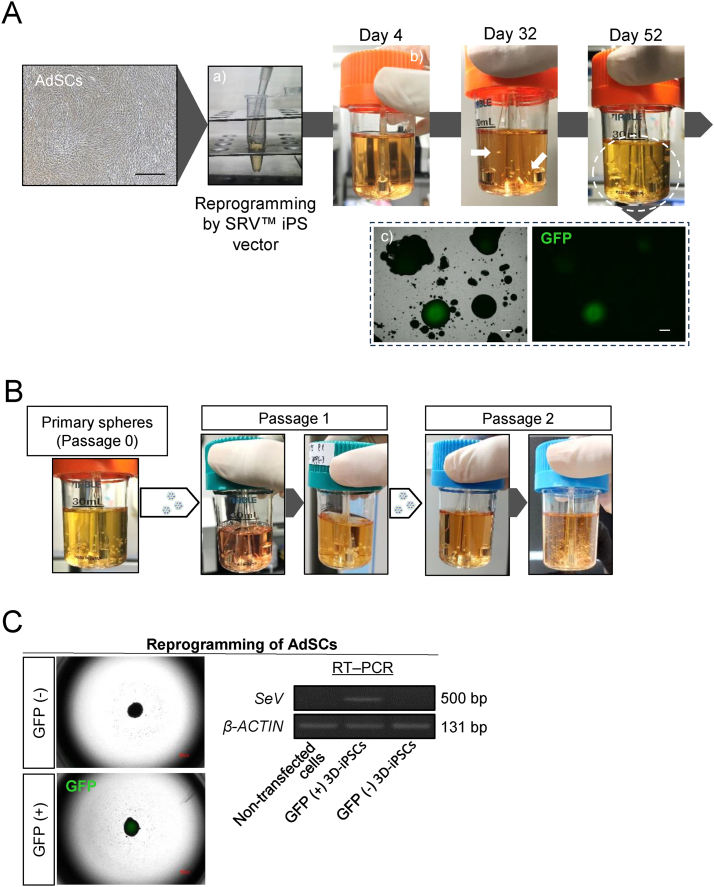
Induced pluripotent stem cells (iPSCs) have immense potential for use in disease modeling, etiological studies, and drug discovery. However, the current workflow for iPSC generation and maintenance poses challenges particularly during the establishment phase when specialized skills are required. Although three-dimensional culture systems offer scalability for maintaining established iPSCs, the enzymatic dissociation step is complex and time-consuming. In this study, a novel approach was developed to address these challenges by enabling iPSC generation, maintenance, and differentiation without the need for two-dimensional culture or enzymatic dissociation. This streamlined method offers a more convenient workflow, reduces variability and labor for technicians, and opens up avenues for advancements in iPSC research and broader applications.

Keywords: 3D culture; Bioreactor; Cell culture; Differentiation; Reprogramming; iPS cells generation.


Conflict of interest statement

The authors have no conflicts of interest to report.

Figures

Fig. 1
Fig. 1
Reprogramming of somatic cells into iPSCs under 3D conditions (A) Representative images depicting the progression of reprogramming under three-dimensional (3D) conditions. Generation of induced pluripotent stem cells (iPSCs) by Sendai virus vectors (SRV™ iPSC Vector). Adipose-derived mesenchymal stem cells (AdSCs) were collected and suspended in a microtube with reprogramming factors for 2 h (a). Cells were then washed and cultured in a single-use bioreactor (b). White arrows indicate primary spheres. Cells were observed by fluorescence microscopy and green fluorescent protein (GFP) expression (Sendai virus vector remnant cells) noted. The sphere cells demonstrated substantial growth (c; white dashed circle) but certain clustered cells exhibited residual expression of GFP (black dashed rectangle). Black/white scale bars = 500 μm. (B) Growth appearance of 3D-iPSCs. Only a few spheres were transferred to the next bioreactor (white arrows). The cells increased in size and number during each passage (dark gray arrows). (C) Left images illustrate GFP-positive or -negative spheres. GFP-negative spheres indicate the absence of Sendai virus (SeV), as shown by quantitative reverse transcription polymerase chain reaction (RT–PCR) image (right panel). AdSCs were donor cells for iPSC reprogramming. β-ACTIN was the housekeeping gene.
Fig. 2
Fig. 2
Characteristics of 3D-iPSCs (A) Alkaline phosphatase (ALP) staining of induced pluripotent stem cells (iPSCs) after adherent culture. Scale bar = 500 μm. (B) Immunostaining of three-dimensional (3D)-iPSC spheres for an undifferentiation state using the markers, TRA-1-60, SSEA4, and OCT4. High magnification images are shown as insets. Scale bar = 100 μm. (C) In vitro differentiation of 3D-iPSCs via embryoid bodies (EBs) expressing markers of the three germ layers. Immunostaining of markers of endoderm (AFP, green), mesoderm (α-SMA, green), and ectoderm (TUBB3, green) layers. Scale bar = 50 μm. (D) Teratoma assay for 3D-iPSCs derived from adipose-derived mesenchymal stem cells (AdSCs). Overall tumor appearance (left) and histological analysis (right). Hematoxylin and eosin staining revealed germ layer derivatives, such as neural tissues (Neu; ectoderm), cartilage (mesoderm), and gut epithelial tissues (Gut; endoderm). Scale bar = 200 μm. (E) Schematic summary of a TaqMan® Human Pluripotent Stem Cell Scorecard™ Panel assessment of 96 genes associated with self-renewal, endoderm, mesoderm, and ectoderm development for 2D- or 3D-iPSCs derived from AdSCs. A heat map and score box plot are shown in the upper panel. Expression plots represent fold change in expression of given genes compared to four samples. 2D-iPSCs and 3D-iPSCs represent undifferentiated states of each iPSC line, and 2D EBs or 3D EBs show self-differentiated states via EB formation from each iPSC line. A graph compares algorithm scores for the expression of given genes (lower panel). (F) Karyotype analysis of 3D-iPSCs at passage 9 using Q-banding. (G) Cell appearance 1 day after thawing (left) and after growth (right). Scale bar = 500 μm.
Fig. 3
Fig. 3
Lineage specification from 3D-iPSCs without enzymatic dissociation steps (A) Schema illustrating the lineage specification procedure from three-dimensional (3D)-induced pluripotent stem cells (iPSCs) under 3D conditions. 3D-iPSC spheres were just transferred to 6-well plates and cultured in differentiation medium on an orbital shaker at 37 °C, 5% CO2 in air. (B) Neural induction and analysis. Morphological changes and cell growth were observed by phase contrast microscopy during the induction process. Scale bar = 500 μm. (C) On day 6, spheres were attached and immunostained for the neural stem cell markers, SOX1 and NESTIN. Hematoxylin-eosin (HE) staining on day 12 revealed rosette-like structures within spheres. Immunostaining showed continued expression of neural stem cell markers. White scale bar = 100 μm, Black scale bar = 500 μm. (D) Cardiac induction and analysis. Morphological changes were observed by phase contrast microscopy during induction. Scale bar = 500 μm. (E) On day 20, differentiated cells were positively stained for the cardiac markers, alpha actin (ACTN2) and cardiac troponin T (TNNT2). Scale bar = 50 μm.
Fig. 4
Fig. 4
Generation and characterization of 3D-iPSCs from PBMCs (A) Workflow of the generation and expansion of peripheral blood mononuclear cell (PBMC)-derived three-dimensional (3D)-induced pluripotent stem cells (iPSCs) under 3D conditions. PBMCs were isolated from human blood and exposed to Sendai virus vector (SRV™ iPS vector). Cells were grown in small-scale (30 mL) spinner bioreactors at 37 °C in a 5% CO2 incubator. White arrows indicate primary spheres. (B) Quantitative reverse transcription polymerase chain reaction (RT–PCR) analysis of Sendai virus (SeV) and a housekeeping gene (β-ACTIN). Sendai virus vector was not detected in Green Fluorescent Protein (GFP)-negative 3D-iPSC cells by RT–PCR. (C) Alkaline phosphatase staining of 3D-iPSCs after adherent culture. Scale bar = 500 μm. (D) Immunostaining of markers for an undifferentiated state, TRA-1-60, SSEA4, and OCT4, in PBMC-derived 3D-iPSCs. Scale bar = 100 μm. (E) Immunostaining of differentiation markers, endoderm (AFP, green), mesoderm (α-SMA, green), and ectoderm (TUBB3, green) for adherent-cultured embryoid bodies (EBs) from PBMC-derived 3D-iPSCs. Scale bar = 50 μm. (F) Tumor formation assay after subcutaneous transplantation of PBMC-derived 3D-iPSCs. Tumor appearance (left). Histological analysis (right). Hematoxylin and eosin staining revealed intestinal tissue (Gut), cartilage tissue (Cartilage), and neural rosettes (Neu). Scale bar = 200 μm. (G) Schematic summary of a TaqMan® Human Pluripotent Stem Cell Scorecard™ Panel assessment of 96 genes associated with self-renewal, endoderm, mesoderm, and ectoderm development for 2D- or 3D-iPSCs derived from PBMCs. iPSCs generated from the same PBMCs using the same Sendai virus vector under 2D conditions (2D-iPSCs) were used as a control. A heat map and score box plot are shown (upper panel). Expression plots represent fold change in expression of given genes compared to four samples. 2D-iPSCs and 3D-iPSCs represent undifferentiated states of each iPSC line, and 2D EBs or 3D EBs showed self-differentiated states via EB formation from 2D- or 3D-iPSCs, respectively. A graph compares algorithm scores for the expression of given genes (lower panel). (H) Karyotype analysis of PBMC-derived 3D-iPSCs at passage six using Q-banding.

Similar articles

Cited by

References

    1. Assou S., Girault N., Plinet M., Bouckenheimer J., Sansac C., Combe M., Mianné J., Bourguignon C., Fieldes M., Ahmed E., et al. Recurrent genetic abnormalities in human pluripotent stem cells: definition and routine detection in culture supernatants by targeted droplet digital PCR. Stem Cell Rep. 2020;14:1–8. doi: 10.1016/J.STEMCR.2019.12.004. - DOI - PMC - PubMed
    1. Bai Q., Ramirez J.M., Becker F., Pantesco V., Lavabre-Bertrand T., Hovatta O., Lemaître J.M., Pellestor F., De Vos J. Temporal analysis of genome alterations induced by single-cell passaging in human embryonic stem cells. Stem Cell Dev. 2015;24:653–662. doi: 10.1089/SCD.2014.0292. - DOI - PMC - PubMed
    1. Castro-Viñuelas R., Sanjurjo-Rodríguez C., Piñeiro-Ramil M., Rodríguez-Fernández S., López-Baltar I., Fuentes-Boquete I., Blanco F.J., Díaz-Prado S. Tips and tricks for successfully culturing and adapting human-induced pluripotent stem cells. Mol Ther Methods Clin Dev. 2021;23:569–581. doi: 10.1016/j.omtm.2021.10.013. - DOI - PMC - PubMed
    1. Fattahi P., Rahimian A., Slama M.Q., Gwon K., Gonzalez-Suarez A.M., Wolf J., Baskaran H., Duffy C.D., Stybayeva G., Peterson Q.P., et al. Core-shell hydrogel microcapsules enable formation of human pluripotent stem cell spheroids and their cultivation in a stirred bioreactor. Sci Rep. 2021;11:7177. doi: 10.1038/s41598-021-85786-2. - DOI - PMC - PubMed
    1. Galvanauskas V., Grincas V., Simutis R., Kagawa Y., Kino-Oka M. Current state and perspectives in modeling and control of human pluripotent stem cell expansion processes in stirred-tank bioreactors. Biotechnol Prog. 2017;33:355–364. doi: 10.1002/btpr.2431. - DOI - PubMed

LinkOut - more resources