Review
. 2024 Mar 10:36:330-357.
doi: 10.1016/j.bioactmat.2024.02.028. eCollection 2024 Jun.

Engineering customized nanovaccines for enhanced cancer immunotherapy

Affiliations
Review

Engineering customized nanovaccines for enhanced cancer immunotherapy

Jinyu Guo et al. Bioact Mater. .

Abstract

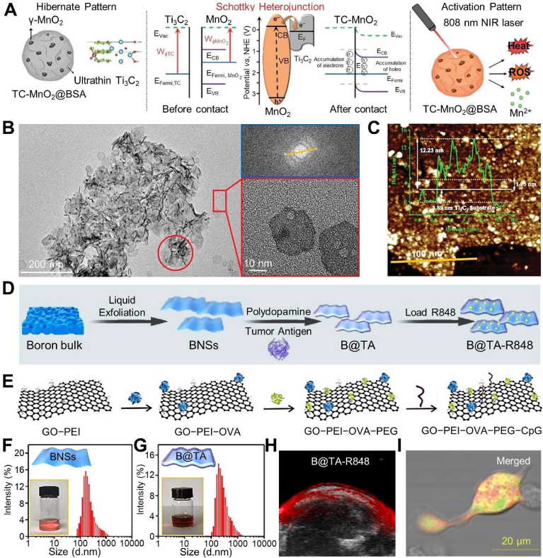
Nanovaccines have gathered significant attention for their potential to elicit tumor-specific immunological responses. Despite notable progress in tumor immunotherapy, nanovaccines still encounter considerable challenges such as low delivery efficiency, limited targeting ability, and suboptimal efficacy. With an aim of addressing these issues, engineering customized nanovaccines through modification or functionalization has emerged as a promising approach. These tailored nanovaccines not only enhance antigen presentation, but also effectively modulate immunosuppression within the tumor microenvironment. Specifically, they are distinguished by their diverse sizes, shapes, charges, structures, and unique physicochemical properties, along with targeting ligands. These features of nanovaccines facilitate lymph node accumulation and activation/regulation of immune cells. This overview of bespoke nanovaccines underscores their potential in both prophylactic and therapeutic applications, offering insights into their future development and role in cancer immunotherapy.

Keywords: Customized structure; Enhanced cancer immunotherapy; Nanovaccines; Prophylactic and therapeutic applications; Tailored-ligand.


Conflict of interest statement

The authors declare no conflict of interest.

Figures

Image 1
Graphical abstract
Scheme 1
Scheme 1
Customized nanovaccines for enhanced cancer immunotherapy.
Fig. 1
Fig. 1
Fabrication processes of hollow structure-based nanovaccines for cancer immunotherapy. Transmission electron microscopy (TEM) images depicting (A) initial H–MnO2, (B) modification of H–MnO2 with an external coating of acrylic acid and the polyelectrolyte poly(allylamine hydrochloride), accompanied by internal loading of doxorubicin (DOX) and Chlorin e6 (Ce6) (H-M-pp/C&D), and (C) subsequent conjugation of modified structure with amino-modified oligonucleotides including CpG, S6-aptamers, and miR-145 (H-M-pp/C&D + 3). (D) Ultraviolet–visible (UV–vis)−near-infrared (NIR) spectra of free Ce6, DOX, H–MnO2, and H-M-pp/C&D. (E) Schematic of synthesis process and various functions of finally prepared nanovaccine. Reproduced with permission [50]. Copyright 2022, American Chemical Society. (F) Synthesis of SiAl NP by successive reverse microemulsion and hydrothermal processes. (G) TEM image and (H) Scanning TEM image of SiAl NP. (I) TF loading efficiencies in SiAl and its alongside incorporation of DOX. (J) OVA release profiles of SiAl@OVA in phosphate-buffered saline solution (PBS) with different pH values. Reproduced with permission [51]. Copyright 2022, Wiley.
Fig. 2
Fig. 2
Fabrication processes of layered structure-based nanovaccines for cancer immunotherapy. (A) Schematic diagram of NIR activated Schottky nanovaccine TC-MnO2@BSA and its mechanisms towards immune effects. (B) TEM images and (C) AFM image and height of TC-MnO2@BSA. Reproduced with permission [57]. Copyright 2023, Wiley. (D) Preparations of personalized nanovaccine of B@TA-R848. Size, dispersity, and photo images of (F) boron nanosheet and (G) its loading TA. (H) Photoacoustic images of tumor site in B@TA-R848 groups at 12 h post injection. Reproduced with permission [58]. Copyright 2021, Walter de Gruyter. (E) Formation illustration of GO/polymer-famulated nanovaccine. (I) Confocal microscope image of BMDCs incubated with GO/polymer-famulated nanovaccine and fluorescein isothiocyanate (FITC). Reproduced with permission [87]. Copyright 2022, Elsevier.
Fig. 3
Fig. 3
Fabrications and applications of micelles in cancer immunotherapy. (A) Schematic diagram of self-assembly micelle, comprising PEG-PE, palmitoylated polypeptide, and MPLA, where hydrophobic components, palmitic acid, and MPLA are integrated into micelle's hydrophobic core. (C) Accumulation of different FITC formulations (FITC; FITC-labeled micelle, F-M; FITC-labeled liposome, F-L) in draining LN (DLN) at indicated time points (axillary LNs are referred as “aLNs” and inguinal LNs are referred as “iLNs”). Reproduced with permission [62]. Copyright 2017, Springer. (B) Schematic illustration of Man-VIPER delivery systems, consisting of disulfide-conjugated OVA antigens and membranolytic melittin (either releasable Man-VIPER-R or non-releasable Man-VIPER-NR). System self-assembles into micelles is endocytosed via mannosylated segments, leading to disassembly in endosomes and subsequent MHC I or MHC II epitope presentation to respective T-cell subsets. (D) Size distributions of non-membranolytic D-melittin-free analogues (Man-AP), Man-VIPER-R and Man-VIPER-NR characterized by dynamic light scattering (DLS; dmean = 27.6 nm, 51.6 nm, 40 nm, respectively). (E) pH-dependent micellization of Man-AP, Man-VIPER-R and Man-VIPER-NR. Reproduced with permission [102]. Copyright 2023, Elsevier.
Fig. 4
Fig. 4
Fabrications and applications of vesicles or nanodisc in cancer immunotherapy. (A) Schematic illustration of personalized nanovaccine by coating adjuvant R837-loaded PLGA NP with calcinetin-expressed Luc-4T1 cell membrane antigens. Reproduced with permission [130]. Copyright 2021, American Chemical Society. (B) Diagram of cMn-MOF@CM nanovaccine. Reproduced with permission [72]. Copyright 2021, Elsevier. (C) Detail mechanism of in situ STING-activating vaccination strategy. (D) Composition of CMM-DiR and functions of each component. (E) Mn2+ release profiles in pH 7.4 and pH 6.8 solutions (H2O2). (F) Expression of proteins on different groups. (G) Western blot for activation of cGAS-STING pathway in DC 2.4 cells with different treatments: Complete reaction liquid (CMM-DiR1), supernatant (CMM-DiRsup), and precipitation (CMM-DiRpre) from co-incubated CMM-DiR and H2O2 solution (pH 6.8). (H) Real-time quantitative polymerase chain reaction analysis for relative expression of cGAS-STING axis in tumor sites of mice with different treatment (n = 5). Reproduced with permission [73]. Copyright 2021, Elsevier. (I) Whole cancer cell derived of TM, followed by incubation with MPLA and styrene-maleic acid to form MPLA-loaded cancer cell membrane nanodisc (CCND/MPLA). (J) Size of cancer cell membrane-based nanodisc (CCND) and CCND/MPLA (n = 3). (K) TEM images of CCND (left) and CCND/MPLA (right) negatively stained with uranyl acetate. (L) Protein profiles of MC38 cell membrane (1), CCND (2), and CCND/MPLA (3) after gel electrophoresis. (M) Western blot probing for tumor antigens in MC38 cell membrane, CCND, and CCND/MPLA. Reproduced with permission [135]. Copyright 2023, American Chemical Society.
Fig. 5
Fig. 5
Nanovaccines for LN-targeted delivery and their enhanced cancer immunotherapy. (A) In situ programming of vaccines via two synergetic nanomedicines, Tu-NPFN and Ln-NPR848. (B) Sodium dodecyl sulfate-polyacrylamide gel electrophoresis of total proteins generated by Tu-NPFN under an alternating magnetic field and proteins captured by Ln-NPR848. Proteins were stained with Coomassie Brilliant Blue. (C) Relative abundances of tumor antigens captured by Ln-NPR848 as determined by liquid chromatography/tandem mass spectrometry. Reproduced with permission [145]. Copyright 2022, American Chemical Society. (D) Schematic illustration of mNV-triggered antitumor immune responses. (G) Representative fluorescence images of DLN for groups of mNV that FAM-labeled CpG (mNVF) at the indicated time points. Reproduced with permission [149]. Copyright 2018, American Chemical Society. (E) Personalized nanoDC that efficiently accumulates in LN after subcutaneous injection, directly stimulating TAA-specific T cells to kill tumor cells. (F) Ex vivo fluorescence image of FITC, FITC-labeled immature DCs membrane-based vaccines (nanoiDCs) or nanoDCs treated LN. Reproduced with permission [185]. Copyright 2022, Wiley.
Fig. 6
Fig. 6
Enhancement of antigen presentation with customized nanovaccines in cancer immunotherapy. (A) Preparation of MOF@cytomembrane. (B) Confocal laser scanning microscopy observation of fusion of DC (green fluorescence from anti-MHC II-FITC antibody) and 4T1 cell (red fluorescence from anti-CD44-APC antibody). Reproduced with permission [153]. Copyright 2019, Springer. (C) Schematic illustration of mannan-decorated pathogen-like polymeric nanoparticle system (MPVax) and mechanism in eliciting antitumor immune responses. MPVax-CpG/Antigen is constructed with PLA-PEI core, loaded with CpG and antigens and shielded with oxidized mannan. MPVax-CpG/Antigen efficiently accumulates in LN by passive drainage or pDC and mannose-binding lectin mediated active transportation. After arriving at CD8+ DC, antigens are presented to CD8+ T cells and results in antigen-specific tumor eradication. Reproduced with permission [154]. Copyright 2022, Elsevier. (D) Formation of size-transformable naAPC. Achieved through self-assembly of copolymer biotin-PEG-b-poly(N-2-hydroxypropyl methacrylamide-g-thiol)-b-poly[2-(dimethylamino) ethyl methacrylate], naAPC encapsulates IL-2 within their aqueous core and features a surface adorned with pMHC monomer and αCD28. (E) Size and structure characterization of naAPC tested by DLS and TEM, respectively. In vitro CD3+CD8+ T cell proliferation by (F) co-incubation of naïve T cells with mature DC 2.4 cells, and co-incubation of mature DC 2.4 cells and dying EG7-OVA cells with (G) T cells; and (H) preactivated T cells by photodynamic therapy (PDT) after treatment with different groups including (1) PBS, (2) naAPC, (3) OVA, (4) NP-OVA, (5) OVA-naAPC, and (6) NP-OVA/naAPC. Mixed cells had PBS or naAPC alone serve as control. (n = 3; **P < 0.01, ***P < 0.001; ns, not significant; one-way ANOVA with multiple comparisons). Reproduced with permission [199]. Copyright 2020, American Association for the Advancement of Science. (I) Schematic illustrations of responsive release and process for triggering anti-tumor immune response of cyclodextrin/PEI-based nanovaccine. Reproduced with permission [201]. Copyright 2023, Wiley.
Fig. 7
Fig. 7
Designs of targeting TAM-specific nanovaccines in cancer immunotherapy. (A) Schematic illustration of macrophage repolarization regulated by peptide hydrogel (Smac-TLR7/8 hydrogel) for overcoming radioresistance. Smac-TLR7/8 peptide self-assembles into nanofibrous hydrogel. Then, (i) after peritumoral injection, Smac-TLR7/8 hydrogel effectively reprograms TAM from M2 type toward M1 type, secreting inflammatory factors and activating anti-tumor functions. (ii) Immunosuppressive TME is rebuilt through tumor infiltrating lymphocytes recruitment, and downregulating Treg cells. (iii) Radioresistance is overcame which further improves anti-tumor efficacy combined with immunotherapy. (B) Percentage of M2-related (F4/80+CD206+) macrophages (n = 5). (C) Protein expression level of NF-κB, TNF-α, inducible isoform of nitric oxide synthase (iNOS), IL-10, arginase 1 in macrophages determined by western blotting. RT: Radiotherapy. Reproduced with permission [166]. Copyright 2022, KeAi Communications Co. Expression level of CD40 (D) and CD206 (E) on bone-marrow-derived macrophage cells that treated with different groups. All the data represented as mean ± SD, n = 3. (F) Application of CpG-Lox-DPNFs vaccines composed of CpG, loxoribine, and co-loaded DNA-polymer hybrid nanocomplex in synergistic reprogramming tumor immune microenvironment for efficient cancer immunotherapy. Reproduced with permission [234]. Copyright 2024, Elsevier.
Fig. 8
Fig. 8
Deductive procedure of FIT nanovaccine for cancer immunotherapy. (A) Preparation process of FIT nanoparticle, mechanism of MDSC regulation, ICD induction process, and dual-imaging medicated enhanced cancer immunotherapy. In vitro cellular experiment with FIT NP, (B) confocal laser scanning microscopy images of CT26 multicellular spheroids treated with different samples for 6 h and stained with FDA (green) and propidium iodide (red). (C) Flow cytometry and (D) qualitative analysis of MDSC in tumors (n = 3; *P < 0.05, **P < 0.01). Reproduced with permission [169]. Copyright 2021, Wiley.

References

    1. McCarthy E.F. The toxins of William B. Coley and the treatment of bone and soft-tissue sarcomas. Lowa Orthop. J. 2006;26:154. - PMC - PubMed
    1. Nam J., Son S., Park K.S., et al. Cancer nanomedicine for combination cancer immunotherapy. Nat. Rev. Mater. 2019;(4):398–414.
    1. Ribas A., Haining W.N., Schumacher T.N.M. When cancer cells become the enablers of an antitumor immune response. Cancer Discov. 2022;(12):2244–2248. - PubMed
    1. Schuster M., Nechansky A., Kircheis R. Cancer immunotherapy. Biotechnol. J.: Healthcare, Nutrition, Technology. 2006;(1):138–147. - PubMed
    1. Saxena M., van der Burg S.H., Melief C.J.M., et al. Therapeutic cancer vaccines. Nat. Rev. Cancer. 2021;(21):360–378. - PubMed

LinkOut - more resources