

Glutaredoxin-1 modulates the NF-κB signaling pathway to activate inducible nitric oxide synthase in experimental necrotizing enterocolitis
- PMID: 38496303
- PMCID: PMC10940916
- DOI: 10.1016/j.omtm.2024.101214
Glutaredoxin-1 modulates the NF-κB signaling pathway to activate inducible nitric oxide synthase in experimental necrotizing enterocolitis
Abstract
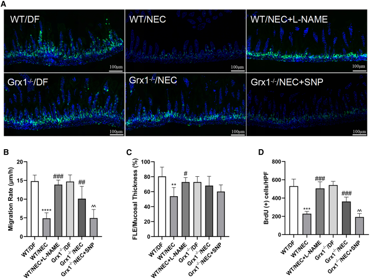
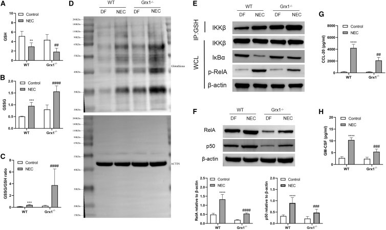
Inducible nitric oxide synthase (iNOS), regulated by nuclear factor kappa B (NF-κB), is crucial for intestinal inflammation and barrier injury in the progression of necrotizing enterocolitis (NEC). The NF-κB pathway is inhibited by S-glutathionylation of inhibitory κB kinase β (IKKβ), which can be restored by glutaredoxin-1 (Grx1). Thus, we aim to explore the role of Grx1 in experimental NEC. Wild-type (WT) and Grx1-knockout (Grx1-/-) mice were treated with an NEC-inducing regimen. Primary intestinal epithelial cells (IECs) were subjected to LPS treatment. The production of iNOS, NO, and inflammation injuries were assessed. NF-κB and involved signaling pathways were also explored. The severity of NEC was attenuated in Grx1-/- mice. Grx1 ablation promoted IKKβ glutathionylation, NF-κB inactivation, and decreased iNOS, NO, and O2·- production in NEC mice. Furthermore, Grx1 ablation restrained proinflammatory cytokines and cell apoptosis, ameliorated intestinal barrier damage, and promoted proliferation in NEC mice. Grx1 ablation protected NEC through iNOS and NO inhibition, which related to S-glutathionylation of IKKβ to inhibit NF-κB signaling. Grx1-related signaling pathways provide a new therapeutic target for NEC.
Keywords: NF-κB; S-glutathionylation; glutaredoxin-1; inducible nitric oxide synthase; necrotizing enterocolitis; oxidative stress.
© 2024 The Authors.
Conflict of interest statement
The authors declare no competing interests.
Figures


Similar articles
-
Contribution of glutaredoxin-1 to S-glutathionylation of endothelial nitric oxide synthase for mesenteric nitric oxide generation in experimental necrotizing enterocolitis.Transl Res. 2017 Oct;188:92-105. doi: 10.1016/j.trsl.2016.01.004. Epub 2016 Jan 18. Transl Res. 2017. PMID: 26845626
-
NF-κB inactivation attenuates the M1 macrophage polarization in experimental necrotizing enterocolitis by glutaredoxin-1 deficiency.Cell Biol Int. 2022 Nov;46(11):1886-1899. doi: 10.1002/cbin.11861. Epub 2022 Jul 23. Cell Biol Int. 2022. PMID: 35870170
-
Activation of the glutaredoxin-1 gene by nuclear factor κB enhances signaling.Free Radic Biol Med. 2011 Sep 15;51(6):1249-57. doi: 10.1016/j.freeradbiomed.2011.06.025. Epub 2011 Jun 27. Free Radic Biol Med. 2011. PMID: 21762778 Free PMC article.
-
Hypoxia-Inducible Factor-1α Modulates the Toll-Like Receptor 4/Nuclear Factor Kappa B Signaling Pathway in Experimental Necrotizing Enterocolitis.Mediators Inflamm. 2024 Dec 17;2024:4811500. doi: 10.1155/mi/4811500. eCollection 2024. Mediators Inflamm. 2024. PMID: 39719983 Free PMC article.
-
Roles of nitric oxide and intestinal microbiota in the pathogenesis of necrotizing enterocolitis.J Pediatr Surg. 2016 Jan;51(1):13-7. doi: 10.1016/j.jpedsurg.2015.10.006. Epub 2015 Oct 22. J Pediatr Surg. 2016. PMID: 26577908 Free PMC article. Review.
Cited by
-
Advancing necrotizing enterocolitis prediction through iterative monitoring.Transl Pediatr. 2024 May 31;13(5):770-783. doi: 10.21037/tp-24-15. Epub 2024 May 20. Transl Pediatr. 2024. PMID: 38840675 Free PMC article.
-
Targeting Inflammation and Oxidative Stress to Improve Outcomes in a TNBS Murine Crohn's Colitis Model.Nanomaterials (Basel). 2024 May 20;14(10):894. doi: 10.3390/nano14100894. Nanomaterials (Basel). 2024. PMID: 38786849 Free PMC article.
References
-
- Ford H.R., Sorrells D.L., Knisely A.S. Inflammatory cytokines, nitric oxide, and necrotizing enterocolitis [J] Semin. Pediatr. Surg. 1996;5:155–159. - PubMed
-
- Yazji I., Sodhi C.P., Lee E.K., Good M., Egan C.E., Afrazi A., Neal M.D., Jia H., Lin J., Ma C., et al. Endothelial TLR4 activation impairs intestinal microcirculatory perfusion in necrotizing enterocolitis via eNOS-NO-nitrite signaling [J] Proc. Natl. Acad. Sci. USA. 2013;110:9451–9456. - PMC - PubMed
LinkOut - more resources
Full Text Sources
