

This is a preprint.
Coronary Microvascular Function Following Severe Preeclampsia
- PMID: 38496439
- PMCID: PMC10942503
- DOI: 10.1101/2024.03.04.24303728
Coronary Microvascular Function Following Severe Preeclampsia
Update in
-
Coronary Microvascular Function Following Severe Preeclampsia.Hypertension. 2024 Jun;81(6):1272-1284. doi: 10.1161/HYPERTENSIONAHA.124.22905. Epub 2024 Apr 2. Hypertension. 2024. PMID: 38563161 Free PMC article.
Abstract
Background: Preeclampsia is a pregnancy-specific hypertensive disorder associated with an imbalance in circulating pro- and anti-angiogenic proteins. Preclinical evidence implicates microvascular dysfunction as a potential mediator of preeclampsia-associated cardiovascular risk.
Methods: Women with singleton pregnancies complicated by severe antepartum-onset preeclampsia and a comparator group with normotensive deliveries underwent cardiac positron emission tomography (PET) within 4 weeks of delivery. A control group of pre-menopausal, non-postpartum women was also included. Myocardial flow reserve (MFR), myocardial blood flow (MBF), and coronary vascular resistance (CVR) were compared across groups. Soluble fms-like tyrosine kinase receptor-1 (sFlt-1) and placental growth factor (PlGF) were measured at imaging.
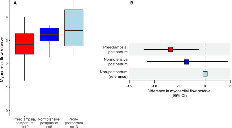
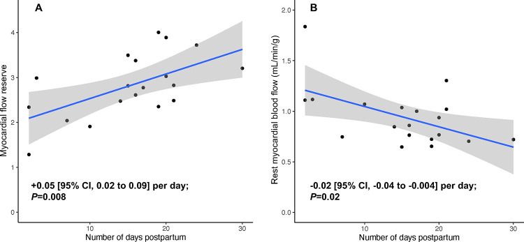
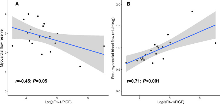
Results: The primary cohort included 19 women with severe preeclampsia (imaged at a mean 16.0 days postpartum), 5 with normotensive pregnancy (mean 14.4 days postpartum), and 13 non-postpartum female controls. Preeclampsia was associated with lower MFR (β=-0.67 [95% CI -1.21 to -0.13]; P=0.016), lower stress MBF (β=-0.68 [95% CI, -1.07 to -0.29] mL/min/g; P=0.001), and higher stress CVR (β=+12.4 [95% CI 6.0 to 18.7] mmHg/mL/min/g; P=0.001) vs. non-postpartum controls. MFR and CVR after normotensive pregnancy were intermediate between preeclamptic and non-postpartum groups. Following preeclampsia, MFR was positively associated with time following delivery (P=0.008). The sFlt-1/PlGF ratio strongly correlated with rest MBF (r=0.71; P<0.001), independent of hemodynamics.
Conclusions: In this exploratory study, we observed reduced coronary microvascular function in the early postpartum period following severe preeclampsia, suggesting that systemic microvascular dysfunction in preeclampsia involves the coronary microcirculation. Further research is needed to establish interventions to mitigate risk of preeclampsia-associated cardiovascular disease.
Keywords: cardiac positron emission tomography; coronary microvascular function; preeclampsia; pregnancy; women’s health.
Figures
References
Publication types
Grants and funding
- T32 HL094301/HL/NHLBI NIH HHS/United States
- K08 HL166687/HL/NHLBI NIH HHS/United States
- K23 HL159276/HL/NHLBI NIH HHS/United States
- R01 HL148565/HL/NHLBI NIH HHS/United States
- R01 HL142711/HL/NHLBI NIH HHS/United States
- R01 HL160003/HL/NHLBI NIH HHS/United States
- R01 HL148050/HL/NHLBI NIH HHS/United States
- R01 HL162960/HL/NHLBI NIH HHS/United States
- R01 EB034586/EB/NIBIB NIH HHS/United States
- R01 HL168889/HL/NHLBI NIH HHS/United States
- K23 HL159243/HL/NHLBI NIH HHS/United States
- K24 HL153669/HL/NHLBI NIH HHS/United States
- K23 HL159279/HL/NHLBI NIH HHS/United States
LinkOut - more resources
Full Text Sources
Miscellaneous