[Preprint]. 2024 Mar 6:2024.03.05.583634.
doi: 10.1101/2024.03.05.583634.

Rare Variable M. tuberculosis Antigens induce predominant Th17 responses in human infection

Affiliations

Rare Variable M. tuberculosis Antigens induce predominant Th17 responses in human infection

Paul Ogongo et al. bioRxiv. .

Abstract

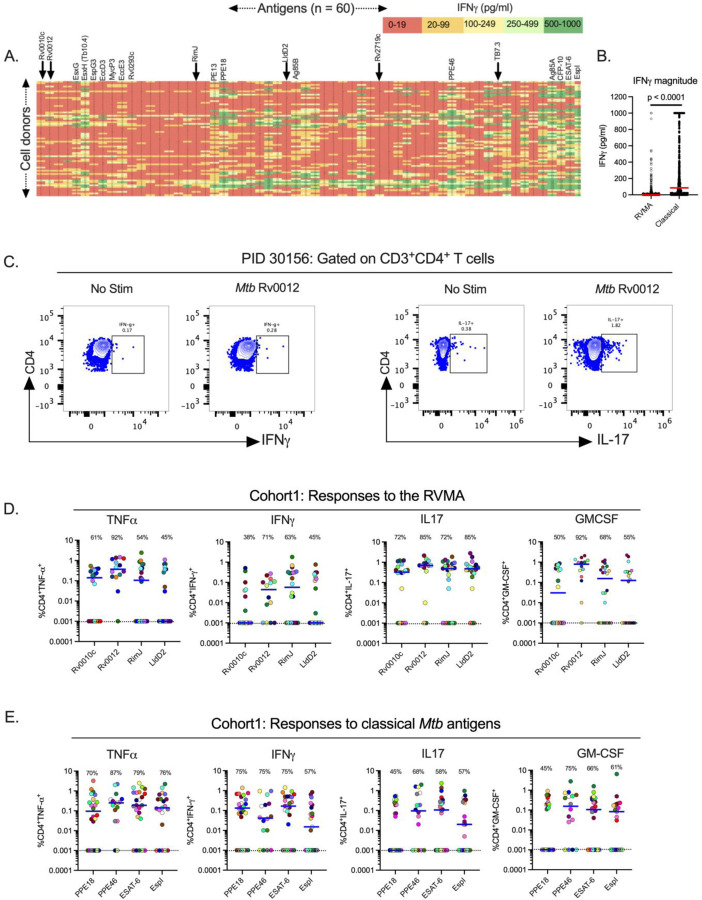
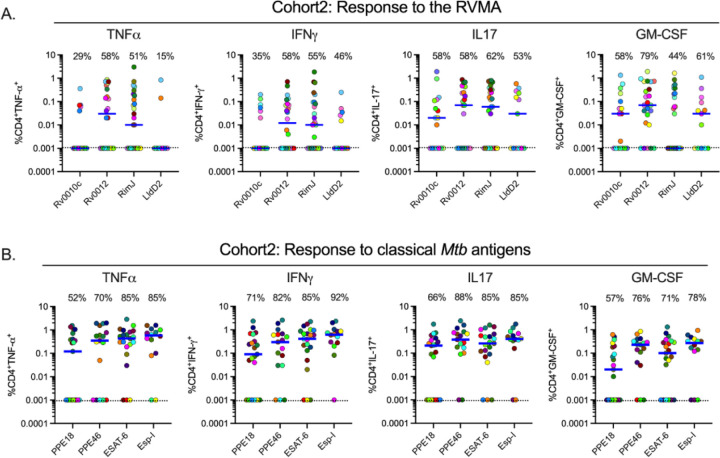
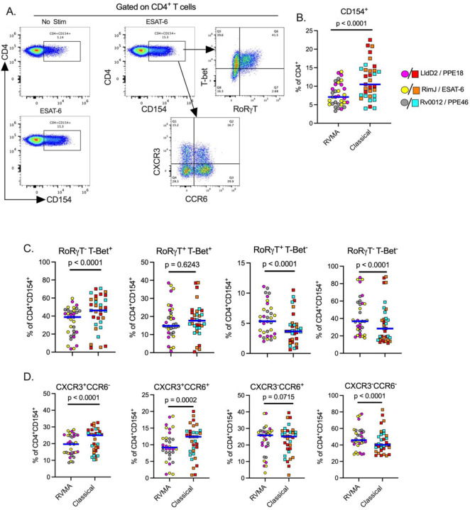
CD4 T cells are essential for immunity to M. tuberculosis (Mtb), and emerging evidence indicates that IL-17-producing Th17 cells contribute to immunity to Mtb. While identifying protective T cell effector functions is important for TB vaccine design, T cell antigen specificity is also likely to be important. To identify antigens that induce protective immunity, we reasoned that as in other pathogens, effective immune recognition drives sequence diversity in individual Mtb antigens. We previously identified Mtb genes under evolutionary diversifying selection pressure whose products we term Rare Variable Mtb Antigens (RVMA). Here, in two distinct human cohorts with recent exposure to TB, we found that RVMA preferentially induce CD4 T cells that express RoRγt and produce IL-17, in contrast to 'classical' Mtb antigens that induce T cells that produce IFNγ. Our results suggest that RVMA can be valuable antigens in vaccines for those already infected with Mtb to amplify existing antigen-specific Th17 responses to prevent TB disease.


Figures

Figure 1:
Figure 1:. Distinct Mtb antigens elicit T cell responses with different functional properties (Cohort 1).
(A) Whole blood samples from QFT+HIV participants in Cohort 1 (KEMRI, Kisumu, Kenya) were stimulated with 60 individual Mtb antigens as overlapping peptides (1μg/ml) for 7 days and supernatants harvested to quantitate IFNγ by ELISA. The antigens are arranged left to right (1–60) according to their positions on the Mtb chromosome, and individual participant samples are arranged in rows. Data presented are results after subtraction of the average of six unstimulated wells as the background response. IFNγ levels are color-coded according to magnitude with red for the lowest and green for the highest responses. Responses greater than the highest standard were assigned the value of the highest standard (1000pg/ml). Selected antigens are highlighted at the top of the heatmap with arrows indicating the RVMA, four of which were studied in more detail using flow cytometry. The identity of all of the antigens ordered in the same fashion is available in (Whatney et al. 2018). (B) Whole blood IFNγ response magnitude of selected classical antigens (Tb10.4 (EsxH), PE13, PPE18, Ag85B, PPE46, Ag85A, CFP-10, ESAT-6 and EspI) compared to the six RVMA (Rv0010c, Rv0012, RimJ, LldD2, Rv2719c and TB7.3), the red horizontal line represents the median response. Statistical significance was determined by Mann-Whitney test. (C) Representative flow cytometry plots comparing CD4 T cells producing IFNg or IL-17 in response to stimulation with one of the RVMA (Rv0012), background response for each cytokine is shown for comparison. (D and E) Cryopreserved peripheral blood mononuclear cells (PBMCs) were stimulated with indicated antigens (2 g/ml) for a total of 20 hours in the presence of Golgi Stop and Golgi Plug and costimulatory antibodies anti-CD28 and anti-CD49d; cytokine production by CD4+ T cells was determined by intracellular cytokine staining and flow cytometry. Data are values after subtraction of unstimulated cells with values lower than the unstimulated control cells indicated below the dotted line (cut-off of positive response). Percent of participants with a detectable response is shown at the top of each antigen plot. Each symbol is a distinct participant, the horizontal blue line indicates the median cytokine response. Results for RVMA are shown in panel C, results for the classical antigens are shown in panel D.
Figure 2:
Figure 2:. Distinct Mtb antigens elicit T cell responses with different functional properties (Cohort 2).
Procedures and analyses were as for Figure 1; the samples were obtained from participants in Cohort 2 (AHRI; Addis Ababa, Ethiopia). Results for RVMA are shown in panel A; results for classical Mtb antigens are shown in panel B.
Figure 3:
Figure 3:. Antigen activated cells express markers of Th17 and Th1 cells upon stimulation with RVMA and classical antigens respectively.
Cryopreserved PBMCs from participants in cohort 1 were stimulated with antigens for a total of 20 hours in the presence of costimulatory antibodies anti-CD28 and anti-CD49d and anti-CD40 blocking antibody, and antigen activated cells identified by CD154 surface expression on CD4+ T cells. (A) Representative flow cytometry plots. (B) CD154 surface expression on CD4+ T cells, CD154+ used to identify antigen activated T cells. (C) Expression of helper T cell lineage defining transcription factors (RORγT = Th17) and (T-bet = Th1) and (D) Expression of chemokine receptors (CCR6 = Th17 marker ) and (CXCR3 = Th1 marker) on antigen activated T cells. Statistics: Wilcoxon matched pairs test.

References

    1. Alwis Ruklanthi de, Williams Katherine L., Schmid Michael A., Lai Chih-Yun, Patel Bhumi, Smith Scott A., Crowe James E., Wang Wei-Kung, Harris Eva, and de Silva Aravinda M. 2014. “Dengue Viruses Are Enhanced by Distinct Populations of Serotype Cross-Reactive Antibodies in Human Immune Sera.” PLoS Pathogens 10 (10): e1004386. 10.1371/journal.ppat.1004386. - DOI - PMC - PubMed
    1. Ardain Amanda, Domingo-Gonzalez Racquel, Das Shibali, Kazer Samuel W., Howard Nicole C., Singh Alveera, Ahmed Mushtaq, et al. 2019. “Group 3 Innate Lymphoid Cells Mediate Early Protective Immunity against Tuberculosis.” Nature, June. 10.1038/s41586-019-1276-2. - DOI - PMC - PubMed
    1. Arlehamn Cecilia Lindestam, Seumois Gregory, Gerasimova Anna, Huang Charlie, Fu Zheng, Yue Xiaojing, Sette Alessandro, Vijayanand Pandurangan, and Peters Bjoern. 2014. “Transcriptional Profile of TB Antigen-Specific T Cells Reveals Novel Multifunctional Features.” Journal of Immunology (Baltimore, Md. : 1950) 193 (6): 2931–40. 10.4049/jimmunol.1401151. - DOI - PMC - PubMed
    1. Barham Morgan S., Whatney Wendy E., Khayumbi Jeremiah, Ongalo Joshua, Sasser Loren E., Campbell Angela, Franczek Meghan, et al. 2020. “Activation-Induced Marker Expression Identifies Mycobacterium Tuberculosis-Specific CD4 T Cells in a Cytokine-Independent Manner in HIV-Infected Individuals with Latent Tuberculosis.” ImmunoHorizons 4 (10): 573–84. 10.4049/immunohorizons.2000051. - DOI - PMC - PubMed
    1. Barnes P. F., Bloch A. B., Davidson P. T., and Snider D. E. 1991. “Tuberculosis in Patients with Human Immunodeficiency Virus Infection.” The New England Journal of Medicine 324 (23): 1644–50. 10.1056/NEJM199106063242307. - DOI - PubMed

Publication types