[Preprint]. 2024 Mar 6:2024.03.06.583756.
doi: 10.1101/2024.03.06.583756.

CD47 is Required for Mesenchymal Progenitor Proliferation and Fracture Repair

Affiliations

CD47 is Required for Mesenchymal Progenitor Proliferation and Fracture Repair

Robert L Zondervan et al. bioRxiv. .

Update in

Abstract

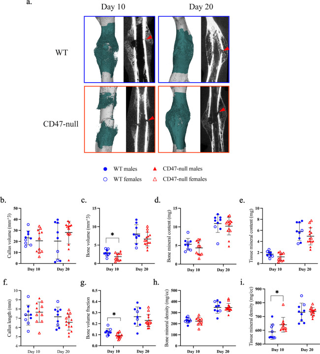
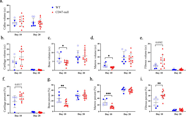
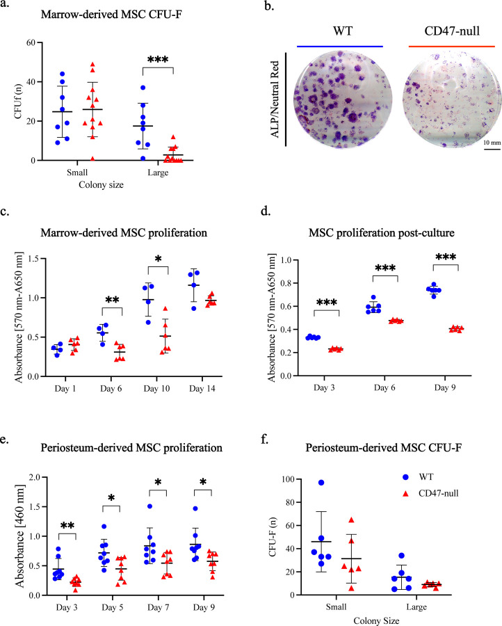
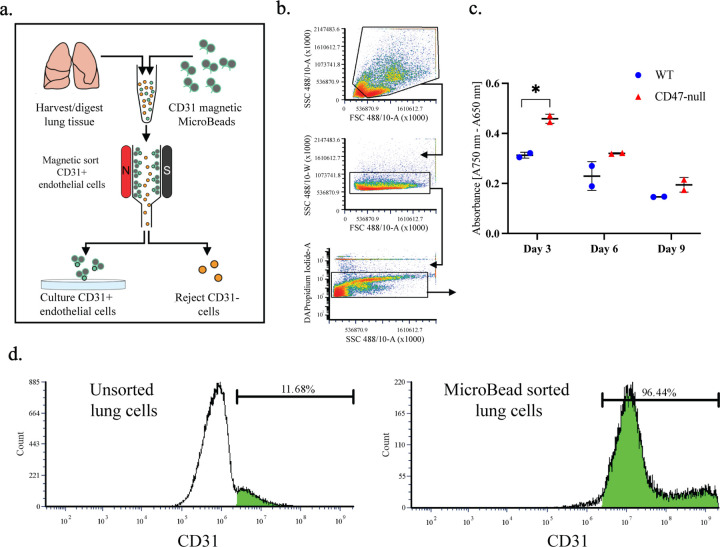
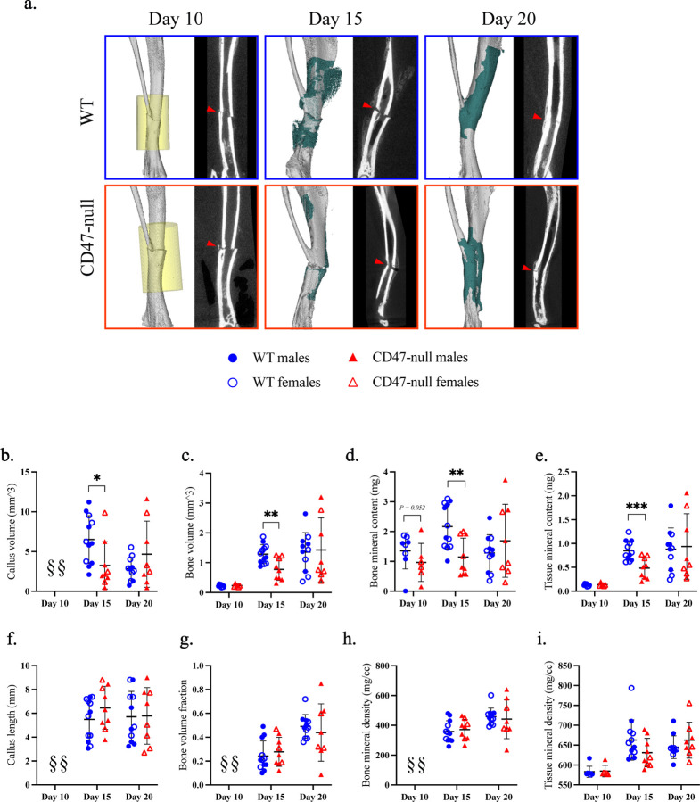
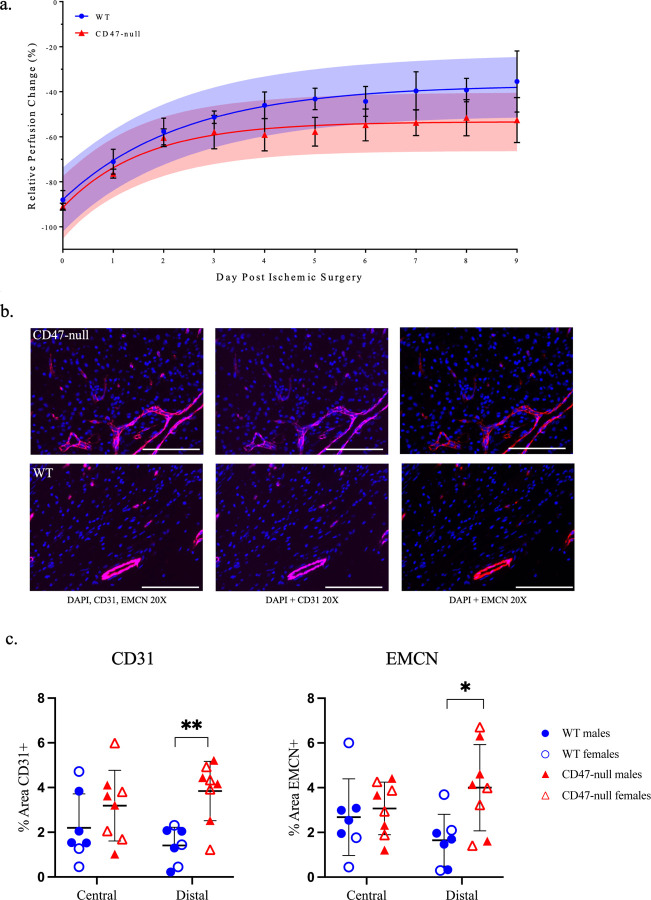
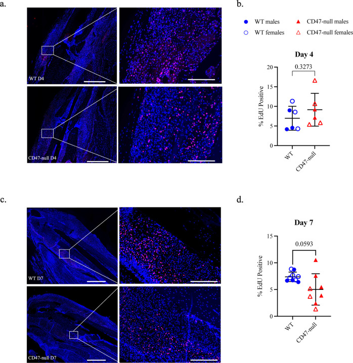
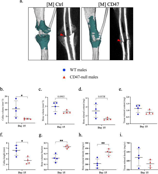
CD47 is a ubiquitous and pleiotropic cell-surface receptor. Disrupting CD47 enhances injury repair in various tissues but the role of CD47 has not been studied in bone injuries. In a murine closed-fracture model, CD47-null mice showed decreased callus bone volume, bone mineral content, and tissue mineral content as assessed by microcomputed tomography 10 days post-fracture, and increased fibrous volume as determined by histology. To understand the cellular basis for this phenotype, mesenchymal progenitors (MSC) were harvested from bone marrow. CD47-null MSC showed decreased large fibroblast colony formation (CFU-F), significantly less proliferation, and fewer cells in S-phase, although osteoblast differentiation was unaffected. However, consistent with prior research, CD47-null endothelial cells showed increased proliferation relative to WT cells. Similarly, in a murine ischemic fracture model, CD47-null mice showed reduced fracture callus bone volume and bone mineral content relative to WT. Consistent with our in vitro results, in vivo EdU labeling showed decreased cell proliferation in the callus of CD47-null mice, while staining for CD31 and endomucin demonstrated increased endothelial cell mass. Finally, WT mice administered a CD47 morpholino, which blocks CD47 protein production, showed a callus phenotype similar to that of non-ischemic and ischemic fractures in CD47-null mice, suggesting the phenotype was not due to developmental changes in the knockout mice. Thus, inhibition of CD47 during bone healing reduces both non-ischemic and ischemic fracture healing, in part, by decreasing MSC proliferation. Furthermore, the increase in endothelial cell proliferation and early blood vessel density caused by CD47 disruption is not sufficient to overcome MSC dysfunction.

Keywords: CD47; Mesenchymal progenitor cell; angiogenesis; callus; fracture healing.


Conflict of interest statement

Conflict of Interests The authors do not have any conflicts of interest to disclose.

Figures

Figure 1.
Figure 1.. CD47-null fractures show reduced bone formation, but increased tissue mineral density
µCT analysis of transverse femoral fracture in WT (n=9) and CD47-null (n=11–14) mice at 10- and 20-days post-fracture. a, Representative 3D reconstructions (white background) and sagittal-plane reconstruction (black background) of WT and CD47-null mice at days 10 and 20 post-fracture. Location of fracture (red arrowhead) is marked on the sagittal reconstructions. Representative 3D reconstructions include callus mineralization (teal). b-i, Callus morphology (mean±SD) at day 10 and 20 post-fracture. *P<0.05, two-sided t test performed at each timepoint.
Figure 2.
Figure 2.. Genetic knockout of the thrombospondin-CD47 axis has minimal effect on late fracture callus composition
Histomorphometry of transverse femoral fractures in WT (n=6–7), and CD47-null (n=11–13) mice at 10- and 20-days post-fracture. a-i, Callus composition quantified through histomorphometry (mean±SD) at day 10 and 20 post-fracture. *P<0.05, **P<0.01, ***P<0.001, two-sided t test performed at each timepoint.
Figure 3.
Figure 3.. CD47 is required for cell colony expansion and proliferation
Marrow and periosteal MSC harvested from the femur and tibia of WT and CD47-null mice. a, CFU-F of WT (n = 8) and CD47-null (n = 12) marrow-derived cells. b, Representative CFU-f plate from WT (left) and CD47-null (right) mice. c, MTT assay of marrow-derived cells MSC from WT (n= 4) and CD47-null (n=4) mice at 1, 6, 10, and 14 days in culture. d, MTT assay of marrow MSC from WT (n= 6) and CD47-null (n=6) after 1 passage at 3, 6, and 9 days in culture. e, CCK8 assay of periosteal MSC from WT (n= 8) and CD47-null (n=8) after 1 passage at 3, 5, 7, and 9 days in culture. f, CFU-F of periosteal MSC from WT (n= 6) and CD47-null (n=6) mice. Mean ±SD, *P<0.05, **P<0.01, ***P<0.001 two-sided t tests.
Figure 4.
Figure 4.. Loss of CD47 decreases percent of MSC in S/G2 phase
Cell cycle analysis of marrow-derived MSC harvested from the femur and tibia of WT (n=5) and D47-null (n=5) mice. a, Representative histograms of WT (left) and CD47-null (right) with cell cycle analysis overlays b, Percent of cells in G1 phase or S/G2. Mean ±SD, *P<0.05, **P<0.01, ***P<0.001, two-sided t test.
Figure 5.
Figure 5.. Loss of CD47 promotes endothelial cell proliferation
a, Schematic of endothelial cell isolation and culture from the lungs of 4-week-old mice. b, Flow cytometry gating scheme for lung cell density (top), double discrimination (middle), and alive/dead (bottom). c, MTT assay of WT and CD47-null endothelial cells after 3, 6, and 9 days in culture. d, Representative histograms of CD31+ lung cells before CD31 MicroBead sorting (left) and after (right). Mean ±SD, *P<0.05, two-sided t test.
Figure 6.
Figure 6.. Genetic knockout of CD47 inhibits early ischemic fracture callus formation
µCT analysis of ischemic tibia fracture callus of WT (n=7–11) and CD47-null (n=6–9) mice at day 10, 15, and 20 post-fracture. a, Representative 3D reconstructions (white background) and sagittal-plane reconstruction (black background) of WT (top row) and CD47-null (bottom row) mice at day 10, 15, and 20 post-fracture. Location of fracture (red arrowhead) is marked on the sagittal reconstructions. Day 10 3D reconstruction includes representative cylindrical ROIs (transparent yellow cylinder) used to calculate callus morphology. Day 15 and 20 representative 3D reconstruction include highlighted callus mineralization (teal). b-i, Callus morphology (mean±SD) at days 10, 15, and 20 post-fracture. *P<0.05, **P<0.01, ***P<0.001, two-sided t tests performed at each timepoint; § no data.
Figure 7.
Figure 7.. Genetic knockout of CD47 limits recovery of whole limb perfusion, but shows local increases in endothelial cells after induced ischemia
Relative perfusion WT and CD47-null mice at days 0–9 post-ischemic surgery using laser doppler flowmetry. a, Perfusion data was fit to a one-phase non-linear curve for WT (fit is the solid blue line with 95% CI in transparent blue; R2=0.7609) and CD47-null (fit is the solid red line with 95% CI in transparent red; R2=0.6223). The rate of recovery was faster, but maximum recovery was significantly lower in CD47-null compared to WT mice (P<0.0001, extra sum-of-squares F test). b, Representative immunofluorescence staining of CD31 and EMCN in CD47-null (n=7) and WT (n=8) ischemic fractures at 20X (Scale bar = 100 µm). c, CD31 quantification in WT and CD47-null ischemic fracture calluses at day 4 post-fracture at peripheral and central regions of the fracture callus. D, EMCN quantification in WT and CD47-null ischemic fracture calluses at day 4 post-fracture at peripheral and central regions. Mean ±SD, *P<0.05, **P<0.01, two-sided t test.
Figure 8.
Figure 8.. Genetic knockout of CD47 reduces cell proliferation in the early fracture callus
EdU expression at 4 and 7 days post ischemic tibia fracture in WT (n=6–9) and CD47-null (n=6–8) mice. a, Representative images of WT and CD47-null fracture calluses at day 4 post-fracture. b, % EdU positive cells in WT and CD47-null fracture callus at 4 days post-fracture. c, Representative images of WT and CD47-null fracture callus at day 7 post-fracture. d, % EdU positive cells in WT and CD47-null fracture calluses at 7 days post-fracture. Two-sided t-test. 10X stitched images, scale bar = 1000 µm. 10X zoomed images, scale bar = 200 µm.
Figure 9.
Figure 9.. Disruption of CD47 using a morpholino inhibits early ischemic fracture callus formation
µCT analysis of ischemic tibia fracture callus of morpholino-control ([M]Ctrl) (n=4) and morpholino-CD47 ([M]CD47) (n=4) at day 15 post-fracture. a, 3D reconstructions (white background with teal mineralized callus highlight) and sagittal-plane reconstruction (black background) of [M]Ctrl (left) and [M]CD47 (right) mice at day 15 post-fracture. Location of fracture (red arrowhead) is marked on the sagittal reconstructions. b-i, Callus morphology (mean±SD) at day 15 post-fracture. *P<0.05, **P<0.01, two-sided t test.

References

    1. Brown E., Hooper L., Ho T. & Gresham H. Integrin-associated protein: a 50-kD plasma membrane antigen physically and functionally associated with integrins. J Cell Biol 111, 2785–2794 (1990). 10.1083/jcb.111.6.2785 - DOI - PMC - PubMed
    1. Lindberg F. P., Gresham H. D., Schwarz E. & Brown E. J. Molecular cloning of integrin-associated protein: an immunoglobulin family member with multiple membrane-spanning domains implicated in alpha v beta 3-dependent ligand binding. J Cell Biol 123, 485–496 (1993). 10.1083/jcb.123.2.485 - DOI - PMC - PubMed
    1. Watanabe 渡邊 直登 N., Takagi 高木 穏香 S., Tominaga 富永 綾 A., Tomita 富田 泰輔 T. & Iwatsubo 岩坪 威 T. Functional Analysis of the Transmembrane Domains of Presenilin 1. J Biol Chem 285, 19738–19746 (2010). 10.1074/jbc.m110.101287 - DOI - PMC - PubMed
    1. Isenberg J. S. et al. Treatment of liver ischemia–reperfusion injury by limiting thrombospondin-1/CD47 signaling. Surgery 144, 752–761 (2008). 10.1016/j.surg.2008.07.009 - DOI - PMC - PubMed
    1. Xiao Z. et al. Attenuation of Ischemia-Reperfusion Injury and Improvement of Survival in Recipients of Steatotic Rat Livers Using CD47 Monoclonal Antibody. Transplantation 100, 1480–1489 (2016). 10.1097/TP.0000000000001186 - DOI - PubMed

Publication types