[Preprint]. 2024 Mar 4:2024.03.04.583369.
doi: 10.1101/2024.03.04.583369.

Comprehensive analysis of CXXX sequence space reveals that S. cerevisiae GGTase-I mainly relies on a2X substrate determinants

Affiliations

Comprehensive analysis of CXXX sequence space reveals that S. cerevisiae GGTase-I mainly relies on a2X substrate determinants

Anushka Sarkar et al. bioRxiv. .

Update in

Abstract

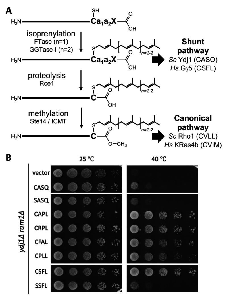
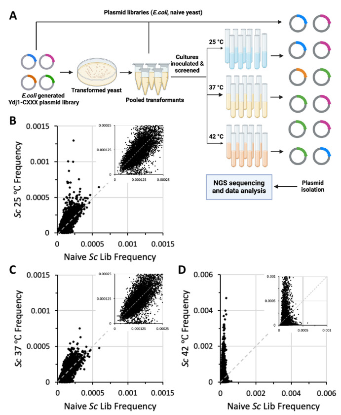
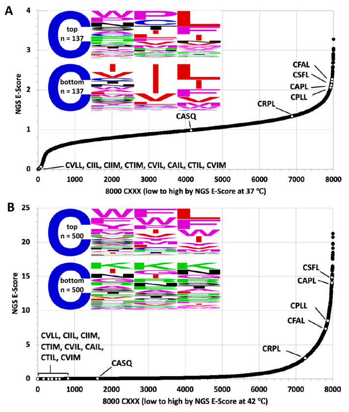
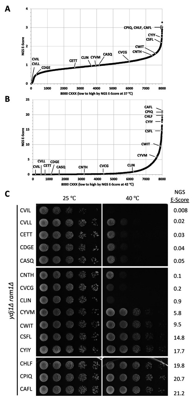
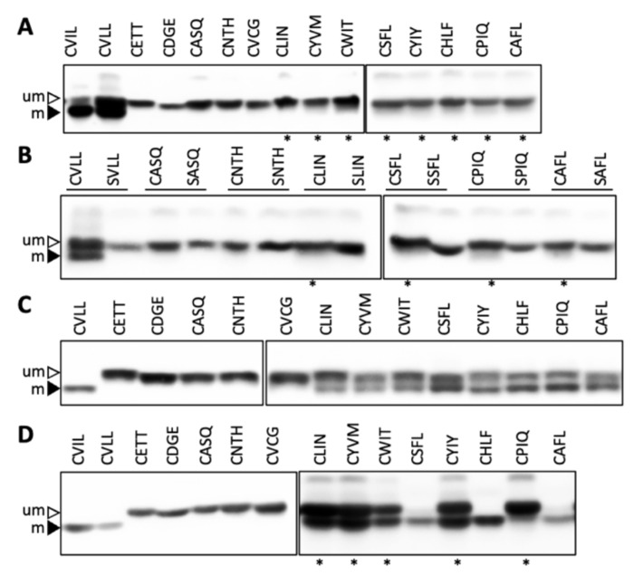
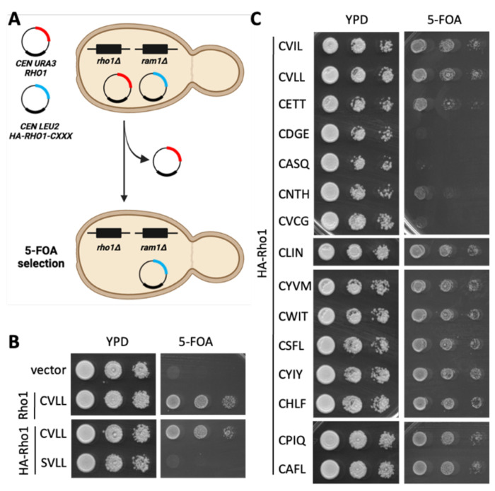
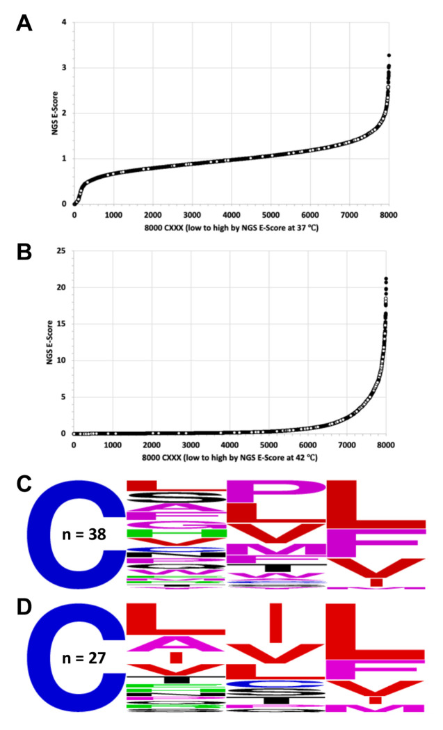
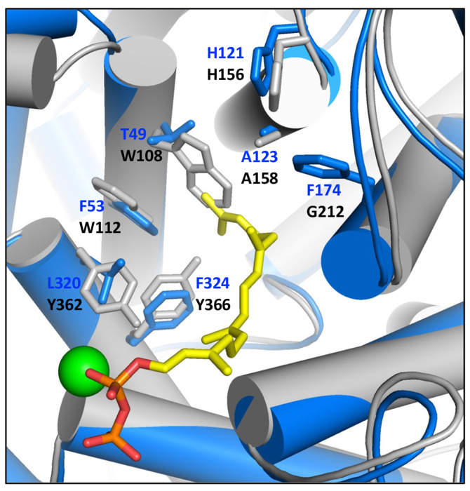
Many proteins undergo a post-translational lipid attachment, which increases their hydrophobicity, thus strengthening their membrane association properties or aiding in protein interactions. Geranylgeranyltransferase-I (GGTase-I) is an enzyme involved in a three-step post-translational modification (PTM) pathway that attaches a 20-carbon lipid group called geranylgeranyl at the carboxy-terminal cysteine of proteins ending in a canonical CaaL motif (C - cysteine, a - aliphatic, L - often leucine, but can be phenylalanine, isoleucine, methionine, or valine). Genetic approaches involving two distinct reporters were employed in this study to assess S. cerevisiae GGTase-I specificity, for which limited data exists, towards all 8000 CXXX combinations. Orthogonal biochemical analyses and structure-based alignments were also performed to better understand the features required for optimal target interaction. These approaches indicate that yeast GGTase-I best modifies the Cxa[L/F/I/M/V] sequence that resembles but is not an exact match for the canonical CaaL motif. We also observed that minor modification of non-canonical sequences is possible. A consistent feature associated with well-modified sequences was the presence of a non-polar a2 residue and a hydrophobic terminal residue, which are features recognized by mammalian GGTase-I. These results thus support that mammalian and yeast GGTase-I exhibit considerable shared specificity.

Keywords: genetic screen; geranylgeranyltransferase-I; next-generation sequencing; target specificity.


Conflict of interest statement

Conflict of interest The authors have declared no competing interests exist.

Figures

Figure 1.
Figure 1.. Geranylgeranylation can be investigated using a Ydj1 reporter.
A) Overview of the CaaX protein modification pathway leading to either shunted (e.g., ScYdj1 or HsGγ5) or canonically modified proteins (e.g., ScRho1, HsKRas4b). B) A Ydj1-based thermotolerance assay can be used to monitor GGTase-I activity. A yeast strain lacking Ydj1 and FTase (yWS2542, ram1Δ ydj1Δ) was transformed with plasmids encoding the indicated Ydj1-CXXX variants. Purified transformants were cultured to saturation in SC-Uracil, and a series of 10-fold dilutions were prepared and spotted onto YPD solid media. Plates were incubated at indicated temperatures for 96 hours (25 °C and 40 °C). Images are representative of results from experiments involving multiple biological and technical replicates.
Figure 2.
Figure 2.. A thermotolerance screen yields an enriched population of Ydj1-CXXX variants.
A) Experimental strategy for probing the entirety of CXXX space that can be modified by yeast GGTase-I. A plasmid library containing all 8000 Ydj1-CXXX combinations was created and transformed into a yeast strain lacking Ydj1 and FTase (yWS2542, ram1Δ ydj1Δ). The transformed colonies were harvested from multiple plates, pooled, and a representative aliquot used to inoculate cultures that were incubated at permissive and restrictive temperatures (25 °C, 37 °C and 42 °C, respectively) until saturation. Plasmids isolated from all populations were sequenced using high throughput methods, and data analyzed to determine the relative frequency of each Ydj1-CXXX variant in each population. Graphic created using BioRender.com. B-D) Plots of frequencies of sequences observed in naïve yeast library relative to those observed after cell expansion under B) 25 °C, C) 37 °C, or D) 42 °C conditions. Inset is a magnification of points near the origin.
Figure 3.
Figure 3.. Temperature effects on the enrichment and de-enrichment of Ydj1-CXXX variants.
Enrichment scores (NGS E-Score) for each of the 8000 Ydj1-CXXX sequences were determined relative to the naïve yeast library for sequences in the A) 37 °C and B) 42 °C yeast libraries. The NGS E-Scores are represented as 2D plots with white dots representing the scores of different sequence sets: i) proteins known or highly suspected to be geranylgeranylated, or not geranylgeranylated (i.e., controls) – Sc Rho1 (CVLL), Sc Rho2 (CIIL), Sc Rho3 (CIIM), Sc Rho4 (CTIM), Sc Rho5 (CVIL), Sc Cdc42 (CAIL), Sc Rsr1 (CTIL), Hs K-Ras4b (CVIM), Sc Ydj1 (CASQ), ii) sequences from the thermotolerance pilot study described in Figure 1B (CRPL, CFAL, CPLL, CAPL), and iii) the sequence derived from Hs Gγ5 (CSFL). The WebLogos associated with each plot reflect an analysis for a subset of sequences. In panel A, the analysis was performed using the 137 highest NGS E-Scores associated with the 37 °C data set (top), and an equivalent number of sequences with the lowest NGS E-Scores <0.2 (bottom). In panel B, the analysis was performed using sequences reflecting the top 500 NGS E-Scores associated with the 42 °C data set (top), and equivalent number of sequences with the lowest NGS E-Scores (bottom).
Figure 4.
Figure 4.. Validation of thermotolerance status for a representative set of Ydj1-CXXX variants.
A-B) The white dots mark the position of 15 sequences that represent a wide distribution of NGS E-Scores on the plots described in Figures 3A and 3B. Included among the sequences representing the Test Set are several predicted to be geranylgeranylated (CSFL, CVLL, and CVIL) or not geranylgeranylated (CASQ) and three that had the highest NGS E-Scores from the 42 °C data set (CHLF, CPIQ, and CAFL). C) Thermotolerance assay results observed for the subset of sequences described in panel A. Sequences were evaluated as described in Figure 1B. NGS E-Scores refer to 42 °C library frequency vs. naïve yeast library frequency values.
Figure 5.
Figure 5.. Evaluation of Ydj1-CXXX variants in the Test Set by gel-shift assay.
A yeast strain lacking Ydj1 and FTase (yWS2542, ram1Δ ydj1Δ) was transformed with plasmids A) from the Test Set or B) matched pairs of sequences encoding either Ydj1-CXXX or its Ydj1-SXXX variant. C-D) The Ydj1-CXXX variants described in panel A were expressed in a yeast strain lacking Ydj1 that overexpresses either C) yeast GGTase-I (yWS4277, ram1Δ ydj1Δ [CEN HIS3 PPGK-RAM2] [CEN LEU2 PPGK-CDC43]) or D) human GGTase-I (yWS3169, ram2Δ ydj1Δ [CEN HIS3 PPGK1-FNTA] [CEN LEU2 PPGK1-PGGT1B]). Cultures were grown at 25 °C or 37 °C (denoted with an *), and total cell lysates were prepared from each transformant condition and analyzed by SDS-PAGE and immunoblot using anti-Ydj1 antibody. Data are representative of two biological replicates. um – unmodified; m – modified.
Figure 6.
Figure 6.. A cell viability assay can distinguish between geranylgeranylated and unmodified Rho1-CXXX variants.
A) Basis for the plasmid loss assay used to assess the function of Rho1-CXXX variants. The yeast strain has chromosomal-disruptions for the FTase β subunit and Rho1 but is viable due to complementation by a URA3-marked plasmid encoding wildtype Rho1 (yWS3761; ram1Δ rho1 [CEN URA3 RHO1]). A second LEU2-marked plasmid encoding a Rho1-CXXX variant is also present (i.e., CEN LEU2 RHO1-CXXX). Upon counterselection with 5-FOA, the URA3-marked plasmid is lost, and yeast will only survive counterselection if the LEU2-marked plasmid encodes a functional Rho1-CXXX variant. Graphic created using BioRender.com and PowerPoint. B-C) Yeast transformed with the indicated CEN LEU2 RHO1-CXXX plasmids were cultured to saturation in SC-Leucine liquid media, and saturated cultures spotted as 10-fold serial dilutions onto 5-FOA and YPD plates. Similar growth patterns on YPD indicate that the serial dilutions were prepared similarly, while growth on 5-FOA indicates the presence of a functional Rho1-CXXX variant. The 15 candidates in panel C are arranged by increasing NGS E-Score (top to bottom).
Figure 7.
Figure 7.. Functional Rho1-CXXX variants can be recovered by the plasmid-loss assay.
A) Experimental strategy for identifying functional Rho1-CXXX variants. A ram1Δ rho1Δ [CEN URA3 RHO1] yeast background was used for co-introduction of a linearized LEU2-based plasmid (CEN LEU2-HA-RHO1-BamHI) and PCR products encoding a library of RHO1-CXXX sequences. Yeast surviving SC-Leucine selection were replica plated onto 5-FOA media, and plasmids recovered and sequenced from 200 yeast colonies surviving counterselection. Graphic created using BioRender.com and PowerPoint. B-D) WebLogo analysis was performed for B) all 112 unique DNA sequences, and sequences conforming to the C) CXX[L/F/I/M/V] consensus and D) CXX[not L/I/F/M/V] consensus.
Figure 8.
Figure 8.. Distribution of NGS-E scores for CXXX sequences identified by Rho1-based screening.
A-B) The 94 CXXX sequences identified by Rho1-based screening are superimposed as white dots on the NGS E-Score plots (A, 37 °C vs. naive yeast library; B, 42 °C vs. naive yeast library) derived from the Ydj1-based screen described in Figures 3A and 3B. C-D). WebLogo analysis of CXXX sequences identified by Rho1-based screening that match the CXX[L/F/I/M/V] consensus and have an NGS E-Score C) >2 or D) <0.5 in the 42 °C data set.
Figure 9.
Figure 9.. Structure-based alignment of rat and yeast GGTase-I β subunits.
The structure of the rat (blue) and yeast (grey) GGTase-I β subunits were derived from PDB 1n4p and an AlphaFold predicted structure, respectively. Active site amino acids are color coded to match the structures; the active site zinc ion is the green sphere; the geranylgeranyl pyrophosphate is indicated in yellow and other colors. Alignment of the structures and an RMSD calculation were performed using the Align function of PyMol.

Similar articles

References

    1. Ashok S., Hildebrandt E. R., Ruiz C. S., Hardgrove D. S., Coreno D. W., Schmidt W. K. and Hougland J. L. (2020). “Protein farnesyltransferase catalyzes unanticipated farnesylation and geranylgeranylation of shortened target sequences.” Biochemistry 59(11): 1149–1162. - PMC - PubMed
    1. Benetka W., Koranda M. and Eisenhaber F. (2006). “Protein Prenylation: An (Almost) Comprehensive Overview on Discovery History, Enzymology, and Significance in Physiology and Disease.” Monatshefte für Chemie / Chemical Monthly 137: 1241–1281.
    1. Berger B. M., Kim J. H., Hildebrandt E. R., Davis I. C., Morgan M. C., Hougland J. L. and Schmidt W. K. (2018). “Protein Isoprenylation in Yeast Targets COOH-Terminal Sequences Not Adhering to the CaaX Consensus.” Genetics 210(4): 1301–1316. - PMC - PubMed
    1. Berger B. M., Yeung W., Goyal A., Zhou Z., Hildebrandt E. R., Kannan N. and Schmidt W. K. (2022). “Functional classification and validation of yeast prenylation motifs using machine learning and genetic reporters.” PLoS One 17(6): e0270128. - PMC - PubMed
    1. Berndt N., Hamilton A. D. and Sebti S. M. (2011). “Targeting protein prenylation for cancer therapy.” Nature Reviews Cancer 11(11): 775–791. - PMC - PubMed

Publication types