[Preprint]. 2024 Mar 9:2024.03.08.582936.
doi: 10.1101/2024.03.08.582936.

Cryo-EM confirms a common fibril fold in the heart of four patients with ATTRwt amyloidosis

Affiliations

Cryo-EM confirms a common fibril fold in the heart of four patients with ATTRwt amyloidosis

Binh An Nguyen et al. bioRxiv. .

Update in

Abstract

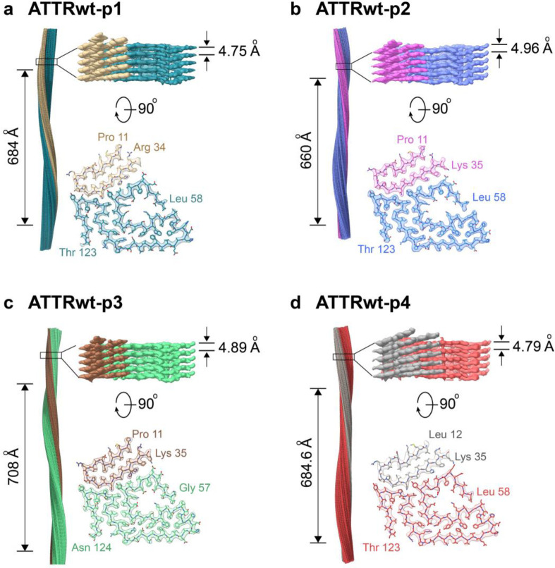
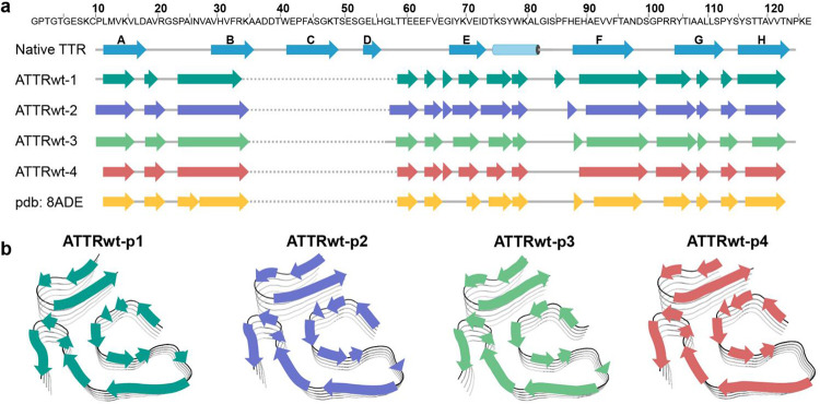
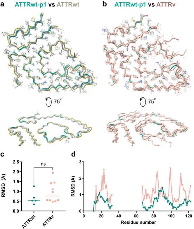
ATTR amyloidosis results from the conversion of transthyretin into amyloid fibrils that deposit in tissues causing organ failure and death. This conversion is facilitated by mutations in ATTRv amyloidosis, or aging in ATTRwt amyloidosis. ATTRv amyloidosis exhibits extreme phenotypic variability, whereas ATTRwt amyloidosis presentation is consistent and predictable. Previously, we found an unprecedented structural variability in cardiac amyloid fibrils from polyneuropathic ATTRv-I84S patients. In contrast, cardiac fibrils from five genotypically-different patients with cardiomyopathy or mixed phenotypes are structurally homogeneous. To understand fibril structure's impact on phenotype, it is necessary to study the fibrils from multiple patients sharing genotype and phenotype. Here we show the cryo-electron microscopy structures of fibrils extracted from four cardiomyopathic ATTRwt amyloidosis patients. Our study confirms that they share identical conformations with minimal structural variability, consistent with their homogenous clinical presentation. Our study contributes to the understanding of ATTR amyloidosis biopathology and calls for further studies.


Figures

Figure 1.
Figure 1.. CryoEM density maps and models of ATTRwt fibrils.
For each patient (a ATTRwt-p1, b ATTRwt-p2, c ATTRwt-p3 and d ATTRwt-p4) figure shows the side view of the reconstructed fibril model with their crossover length (left), the closeup side view of the map with its helical rise (top right), and the top view of a single fibril layer with its model consisting N-terminal fragment (Pro 11/Leu 12 to Arg 34/Lys 35) and C-terminal fragment (Gly 57/Leu 58 to Thr 123/Asn 124) (bottom right.) The color coding is consistent in the cross-sectional model and in the side view of the 3D map.
Figure 2.
Figure 2.. Secondary structure of ATTRwt fibrils.
a Primary sequence of wild-type transthyretin (top) with secondary structures of native-folded transthyretin (PDB 4TLT) and fibrils from four ATTRwt amyloidosis patients determined in this study (PDB codes 8E7D, 8G9R, 8GBR, and 8E7H) and previous study (PDB 8ADE). b Schematic view of the secondary structure on the fibril models of four ATTRwt fibrils.
Figure 3.
Figure 3.. Fibril stability and composition in ATTRwt-p1 amyloidosis.
a Representation of solvation energies per residue estimated from ATTRwt-p1 fibril structures determined in this study. Residues are colored from favorable (red, −2.5 kcal/mol) to unfavorable stabilization energy (blue, 2.5 kcal/mol). Scale, 10 Å. b Schematic view of ATTRwt-p1 fibril structures with residue composition. Residues are color-coded by amino acid category, as labeled. Scale, 10 Å.
Figure 4.
Figure 4.. Backbone comparative analysis of ATTR fibril structures.
a Structure backbone alignment of ATTRwt fibrils with top view (top) and side view (bottom). ATTRwt-p1, in green, is used as a reference, ATTRwt-p2 is in light yellow and three other ATTRwt(s) are in light grey (two from this study and a published ATTRwt PDB 8ADE). b Structure backbone alignment of ATTRwt-p1 fibrils, in green; and other published ATTRv structures, in light pink (PDB codes 8E7E, 8E7J, 8E7I, 8TDN, 8TDO, 7OB4, 8PKG, 8PKE, 6SDZ, 8PKF). c Overall root mean square deviation (RMSD, only comparing Ca in Å) analysis of the structures included in (a) (ATTRwt) and (b) (ATTRv only) using GESAMT Lines indicate mean/median values. d Dot plot of the RMSD analysis (only comparing Ca in Å) per residue from the same groups as in (c). Each dot represents the average RMSD of comparing to a consensus sequence calculated by GESAMT for each group.
Figure 5.
Figure 5.. Potential aggregation pathway of transthyretin into ATTR fibrils with minimal structural alteration.
a, Tetrameric transthyretin (PDB 4TLT). b, Monomer unit after dissociating from tetrameric form. c, Strands C and D flip away to expose strand A and B. d, Strands A and B form hairpin structure while strand E and helix unwind to shape into the core of the polar channel. e, Strand G and H rotate to approximately 135° to strand F, so stand F is now free to interact with the back of the polar channel though the formation of an aromatic pocket. f, The loop region of strand H and the C-terminal part of strand G unfolds to form the triquetra that connects to the N-terminal hairpin and the back of the polar channel. g, Strand H rotates to break the β-sheet hydrogen bonds with strand G and create new interaction at the back of strand G. h, Top view of a single layer of the mature ATTR fibril. Color codes represent different regions of the ATTR fibril and monomeric transthyretin. Red, N-terminal hairpin. Purple, unstructured region encompassing strands C and D, which connects the N-terminal to the C-terminal fragments of the ATTR fibril. Pink, the loop between strand D and E that forms the gate of the polar channel. Green, strands E, F, G, and H that form the rest of the C-terminal fragment of the ATTR fibril.

Similar articles

References

    1. Tschöpe C. & Elsanhoury A. Treatment of Transthyretin Amyloid Cardiomyopathy: The Current Options, the Future, and the Challenges. J Clin Med 11(2022). - PMC - PubMed
    1. Buxbaum J.N. & Reixach N. Transthyretin: the servant of many masters. Cell Mol Life Sci 66, 3095–3101 (2009). - PMC - PubMed
    1. Bezerra F., Saraiva M.J. & Almeida M.R. Modulation of the Mechanisms Driving Transthyretin Amyloidosis. Front Mol Neurosci 13, 592644 (2020). - PMC - PubMed
    1. González-López E., et al. Wild-type transthyretin amyloidosis as a cause of heart failure with preserved ejection fraction. European Heart Journal 36, 2585–2594 (2015). - PubMed
    1. Narotsky D.L., Castano A., Weinsaft J.W., Bokhari S. & Maurer M.S. Wild-Type Transthyretin Cardiac Amyloidosis: Novel Insights From Advanced Imaging. Can J Cardiol 32, 1166.e1161–1166.e1110 (2016). - PMC - PubMed

Publication types