

This is a preprint.
Meningeal contrast enhancement in multiple sclerosis: assessment of field strength, acquisition delay, and clinical relevance
- PMID: 38496664
- PMCID: PMC10942534
- DOI: 10.1101/2024.03.04.24303491
Meningeal contrast enhancement in multiple sclerosis: assessment of field strength, acquisition delay, and clinical relevance
Update in
-
Meningeal contrast enhancement in multiple sclerosis: Assessment of field strength, acquisition delay, and clinical relevance.PLoS One. 2024 May 29;19(5):e0300298. doi: 10.1371/journal.pone.0300298. eCollection 2024. PLoS One. 2024. PMID: 38809920 Free PMC article.
Abstract
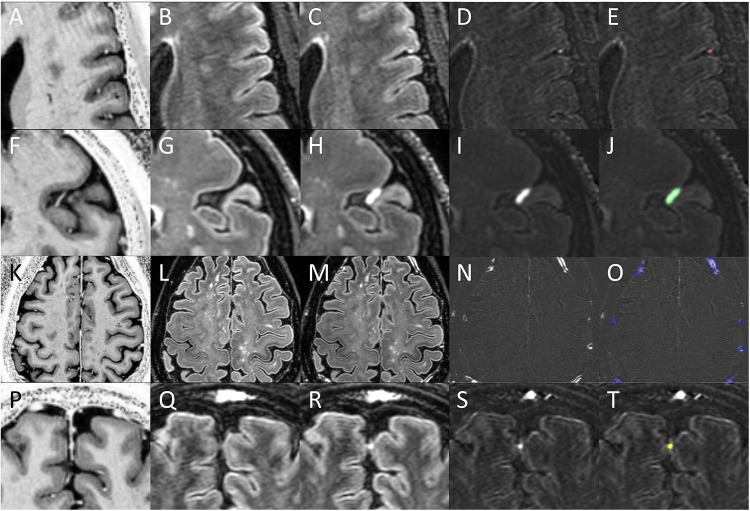
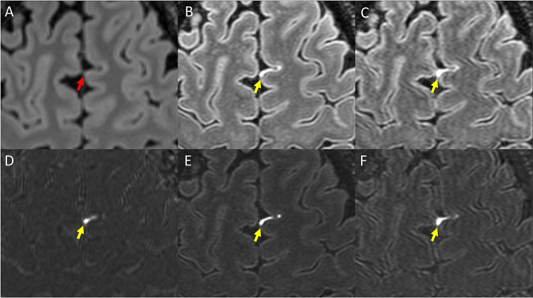
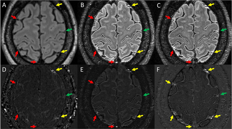
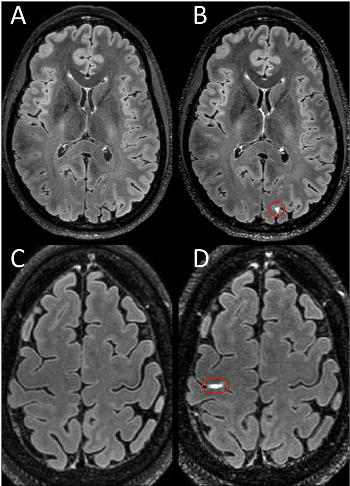
Background/purpose: Leptomeningeal enhancement (LME) on post-contrast FLAIR is described as a potential biomarker of meningeal inflammation in multiple sclerosis (MS). Here we report a comprehensive assessment of the impact of MRI field strength and acquisition timing on meningeal contrast enhancement (MCE).
Methods: This was a cross-sectional, observational study of 95 participants with MS and 17 healthy controls (HC) subjects. Each participant underwent an MRI of the brain on both a 7 Tesla (7T) and 3 Tesla (3T) MRI scanner. 7T protocols included a FLAIR image before, soon after (Gd+ Early 7T FLAIR), and 23 minutes after gadolinium (Gd+ Delayed 7T FLAIR). 3T protocol included FLAIR before and 21 minutes after gadolinium (Gd+ Delayed 3T FLAIR).
Results: LME was seen in 23.3% of participants with MS on Gd+ Delayed 3T FLAIR, 47.4% on Gd+ Early 7T FLAIR (p = 0.002) and 57.9% on Gd+ Delayed 7T FLAIR (p < 0.001 and p = 0.008, respectively). The count and volume of LME, leptomeningeal and paravascular enhancement (LMPE), and paravascular and dural enhancement (PDE) were all highest for Gd+ Delayed 7T FLAIR and lowest for Gd+ Delayed 3T FLAIR. Non-significant trends were seen for higher proportion, counts, and volumes for LME and PDE in MS compared to HCs. The rate of LMPE was different between MS and HCs on Gd+ Delayed 7T FLAIR (98.9% vs 82.4%, p = 0.003). MS participants with LME on Gd+ Delayed 7T FLAIR were older (47.6 (10.6) years) than those without (42.0 (9.7), p = 0.008).
Conclusion: 7T MRI and a delay after contrast injection increased sensitivity for all forms of MCE. However, the lack of difference between groups for LME and its association with age calls into question its relevance as a biomarker of meningeal inflammation in MS.
Figures
References
-
- Howell OW, Reeves CA, Nicholas R, Carassiti D, Radotra B, Gentleman SM, et al. Meningeal inflammation is widespread and linked to cortical pathology in multiple sclerosis. Brain. 2011;134(Pt 9):2755–71. - PubMed
-
- Magliozzi R, Howell OW, Reeves C, Roncaroli F, Nicholas R, Serafini B, et al. A Gradient of neuronal loss and meningeal inflammation in multiple sclerosis. Ann Neurol. 2010;68(4):477–93. - PubMed
-
- Vercellino M, Costantini G, Cogoni M, Lequio L, Sciortino P, De Negri F, et al. Association of MRI leptomeningeal enhancement with disability worsening in progressive multiple sclerosis: A clinical and post-mortem study. Mult Scler. 2023:13524585231199031. - PubMed
-
- Magliozzi R, Howell O, Vora A, Serafini B, Nicholas R, Puopolo M, et al. Meningeal B-cell follicles in secondary progressive multiple sclerosis associate with early onset of disease and severe cortical pathology. Brain. 2007;130(Pt 4):1089–104. - PubMed
-
- Eisele P, Griebe M, Szabo K, Wolf ME, Alonso A, Engelhardt B, et al. Investigation of leptomeningeal enhancement in MS: a postcontrast FLAIR MRI study. Neurology. 2015;84(8):770–5. - PubMed
Publication types
Grants and funding
LinkOut - more resources
Full Text Sources