

Case Reports
doi: 10.1016/j.hrcr.2023.12.001.
eCollection 2024 Mar.
Distinct single spiky component of local abnormal ventricular activity and accurate identification of origin of premature ventricular complexes arising from left ventricular summit
Affiliations
- PMID: 38496738
- PMCID: PMC10943545
- DOI: 10.1016/j.hrcr.2023.12.001
Item in Clipboard
Case Reports
Distinct single spiky component of local abnormal ventricular activity and accurate identification of origin of premature ventricular complexes arising from left ventricular summit
HeartRhythm Case Rep.
.
No abstract available
Keywords: 2F microelectrode catheter; 3-D mapping system; Left ventricular summit; Local abnormal ventricular activity; Premature ventricular complex.
Figures
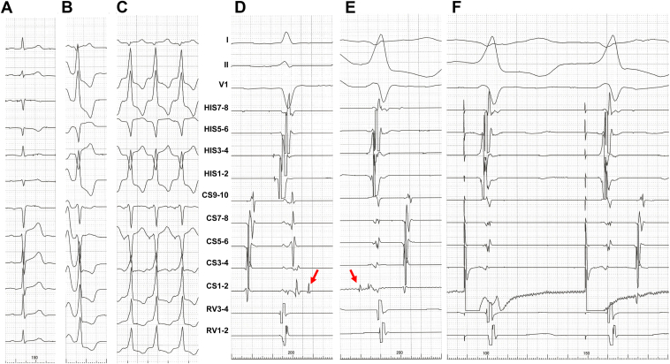
A–C: Twelve-lead electrocardiogram (ECG) during sinus rhythm (A), clinical premature ventricular complex (PVC) (B), and electrical bipolar stimulation from the earliest site of distal bipolar microelectrodes (CS 1-2) with excellent match for the QRS complex of clinical PVCs (C). D–F: Surface ECG (I, II, and V1) and intracardiac electrograms during sinus rhythm (D), clinical PVC (E), and electrical stimulation from CS 1-2 (F). Red arrows indicate a single spiky component of local abnormal ventricular activity, late potential during sinus rhythm (D) and prepotential during clinical PVC (E). CS = coronary sinus; HIS = His bundle electrogram; RV = right ventricular apex.
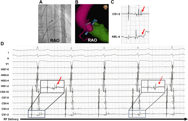
A: Fluoroscopic image. B: Impedance-based electroanatomical mapping images. C: Intracardiac electrogram at CS 1-2 and ABL-d. The red arrows show the delayed single spiky component of the local abnormal ventricular activity (LAVA) at CS 1-2 and far-field delayed potential of the LAVA at ABL-d during sinus rhythm, respectively. D: Intracardiac electrogram taken immediately after radiofrequency application. The solid and dotted black rectangles indicate the potential of the distal coronary sinus microelectrodes during sinus rhythm and at the 4 beats after radiofrequency (RF) application, respectively. The solid and dotted red arrows indicate the single spiky component of LAVA and its disappearance at the 4 beats after RF application, respectively. ABL-d = distal site of the ablation catheter; HIS = His bundle electrogram; RAO = right anterior oblique 30° view; RV = right ventricular apex.
Similar articles
-
Intramyocardial mapping of ventricular premature depolarizations via septal venous perforators: Differentiating the superior intraseptal region from left ventricular summit origins.Heart Rhythm. 2022 Sep;19(9):1475-1483. doi: 10.1016/j.hrthm.2022.03.004. Epub 2022 Mar 10. Heart Rhythm. 2022. PMID: 35278700
-
Approach selection of radiofrequency catheter ablation for ventricular arrhythmias originating from the left ventricular summit: potential relevance of Pseudo Delta wave, Intrinsicoid deflection time, maximal deflection index.BMC Cardiovasc Disord. 2017 May 30;17(1):140. doi: 10.1186/s12872-017-0575-5. BMC Cardiovasc Disord. 2017. PMID: 28558750 Free PMC article.
-
Surgical Mapping and Ablation in the Left Ventricular Summit Guided by Presurgery Pericardial Mapping.J Innov Card Rhythm Manag. 2019 Mar 15;10(3):3582-3587. doi: 10.19102/icrm.2019.100306. eCollection 2019 Mar. J Innov Card Rhythm Manag. 2019. PMID: 32477721 Free PMC article.
-
Catheter Ablation of Ventricular Arrhythmias Arising from the Left Ventricular Summit.Card Electrophysiol Clin. 2016 Mar;8(1):99-107. doi: 10.1016/j.ccep.2015.10.011. Epub 2016 Jan 8. Card Electrophysiol Clin. 2016. PMID: 26920177 Review.
-
A Comprehensive Review of Left Ventricular Summit Ventricular Arrhythmias.J Tehran Heart Cent. 2022 Jul;17(3):91-102. doi: 10.18502/jthc.v17i3.10841. J Tehran Heart Cent. 2022. PMID: 37252083 Free PMC article. Review.
References
-
- Yamada T., Yoshida N., Doppalapudi H., Litovsky S.H., McElderry H.T., Kay G.N. Efficacy of an anatomical approach in radiofrequency catheter ablation of idiopathic ventricular arrhythmias originating from the left ventricular outflow tract. Circ Arrhythm Electrophysiol. 2017;10 - PubMed
-
- Jais P., Maury P., Khairy P., et al. Elimination of local abnormal ventricular activities: a new end point for substrate modification in patients with scar-related ventricular tachycardia. Circulation. 2012;125:2184–2196. - PubMed
-
- Komatsu Y., Daly M., Sacher F., et al. Electrophysiologic characterization of local abnormal ventricular activities in postinfarction ventricular tachycardia with respect to their anatomic location. Heart Rhythm. 2013;10:1630–1637. - PubMed
-
- Komatsu Y., Daly M., Sacher F., et al. Endocardial ablation to eliminate epicardial arrhythmia substrate in scar-related ventricular tachycardia. J Am Coll Cardiol. 2014;63:1416–1426. - PubMed
-
- Santangeli P., Marchlinski F.E., Zado E.S., et al. Percutaneous epicardial ablation of ventricular arrhythmias arising from the left ventricular summit: outcomes and electrocardiogram correlates of success. Circ Arrhythm Electrophysiol. 2015;8:337–343. - PubMed
Publication types
LinkOut - more resources
Full Text Sources
