. 2024 Mar 18;8(4):e0399.
doi: 10.1097/HC9.0000000000000399. eCollection 2024 Apr 1.

ER stress promotes mitochondrial calcium overload and activates the ROS/NLRP3 axis to mediate fatty liver ischemic injury

Affiliations

ER stress promotes mitochondrial calcium overload and activates the ROS/NLRP3 axis to mediate fatty liver ischemic injury

Fei Li et al. Hepatol Commun. .

Abstract

Background: Fatty livers are widely accepted as marginal donors for liver transplantation but are more susceptible to liver ischemia and reperfusion (IR) injury. Increased macrophage-related inflammation plays an important role in the aggravation of fatty liver IR injury. Here, we investigate the precise mechanism by which endoplasmic reticulum (ER) stress activates macrophage NOD-like receptor thermal protein domain-associated protein 3 (NLRP3) signaling by regulating mitochondrial calcium overload in fatty liver IR.

Methods: Control- and high-fat diet-fed mice were subjected to a partial liver IR model. The ER stress, mitochondrial calcium levels, and NLRP3 signaling pathway in macrophages were analyzed.

Results: Liver steatosis exacerbated liver inflammation and IR injury and enhanced NLRP3 activation in macrophages. Myeloid NLRP3 deficiency attenuated intrahepatic inflammation and fatty liver injury following IR. Mechanistically, increased ER stress and mitochondrial calcium overload were observed in macrophages obtained from mouse fatty livers after IR. Suppression of ER stress by tauroursodeoxycholic acid effectively downregulated mitochondrial calcium accumulation and suppressed NLRP3 activation in macrophages, leading to decreased inflammatory IR injury in fatty livers. Moreover, Xestospongin-C-mediated inhibition of mitochondrial calcium influx decreased reactive oxygen species (ROS) expression in macrophages after IR. Scavenging of mitochondrial ROS by mito-TEMPO suppressed macrophage NLRP3 activation and IR injury in fatty livers, indicating that excessive mitochondrial ROS production was responsible for macrophage NLRP3 activation induced by mitochondrial calcium overload. Patients with fatty liver also exhibited upregulated activation of NLRP3 and the ER stress signaling pathway after IR.

Conclusions: Our findings suggest that ER stress promotes mitochondrial calcium overload to activate ROS/NLRP3 signaling pathways within macrophages during IR-stimulated inflammatory responses associated with fatty livers.


Conflict of interest statement

The authors have no conflicts to report.

Figures

None
Graphical abstract
FIGURE 1
FIGURE 1
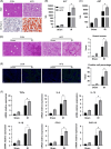
Fatty liver aggravated hepatic ischemia-reperfusion injury. Male C57BL/6 wild-type mice were fed with a normal chow or high-fat diet. Liver tissues were collected after 5 months of feeding. (A) H&E and Oil Red O staining of liver sections. Control and HFD-fed mice were treated with liver IR model or sham procedure. Liver tissues and blood samples were collected at 6 hours after reperfusion. (B, C) Serum ALT and AST levels. (D) H&E staining of liver sections; Suzuki scores based on H&E. (E) TUNEL staining of liver sections; percentage of positive cells were evaluated by ImageJ software. (F) Gene expression of TNF-α, IL-6, IL-18, IL-1β, CCL2, and CXCL10 in liver tissues measured by qRT-PCR. n=4 mice/group. Values were presented as the mean±SD. *p<0.05. Abbreviations: ALT, alanine aminotransferase; AST, aspartate aminotransferase; CCL2, C-C motif chemokine ligand 2; CXCL10, CXCchemokineligand-10; H&E, hematoxylin-eosin; HFD, high-fat diet; IR, ischemia and reperfusion; qRT, quantitative Real-Time; TUNEL, terminal deoxynucleotidyl transferase biotin-dUTP nick end labeling.
FIGURE 2
FIGURE 2
Fatty liver activated NLRP3 in intrahepatic macrophages. Control and HFD-fed mice were treated with liver IR model or sham procedure. Liver tissues and intrahepatic macrophages were collected at 6 hours after reperfusion. (A) Immunofluorescence staining of DAPI (blue) and F4/80 (red) in liver tissues; percentage of F4/80-positive cells was evaluated by ImageJ software. (B) Immunofluorescence staining of DAPI (blue), F4/80 (red), and NLRP3 (green) in liver tissues; percentage of F4/80 and NLRP3-positive cells was evaluated by ImageJ software. (C) Western blot analysis of NLRP3, C-caspase-1, Pro-caspase-1, IL-1β, IL-18, and GAPDH in isolated macrophages. n=4 mice/group. Values were presented as the mean±SD. *p<0.05. Abbreviations: GAPDH, glyceraldehyde 3-phosphate dehydrogenase; HFD, high-fat diet; IR, ischemia and reperfusion; NLRP3, NOD-like receptor thermal protein domain–associated protein 3.
FIGURE 3
FIGURE 3
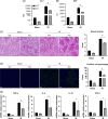
NLRP3 myeloid knockout alleviates IR injury in fatty liver. WT and NLRP3 MKO mice were fed with a high-fat diet, then subjected to liver IR model or sham procedure. Liver tissues and blood samples were collected at 6 hours after reperfusion. (A, B) Serum ALT and AST levels. (C) H&E staining of liver sections; Suzuki scores based on H&E. (D) TUNEL staining of liver sections; percentage of positive cells was evaluated by ImageJ software. (E) Gene expression of TNF-α, IL-6, IL-18, and CCL2 in liver tissues measured by qRT-PCR. n=4 mice/group. Values were presented as the mean±SD. *p<0.05. Abbreviations: ALT, alanine aminotransferase; AST, aspartate aminotransferase; CCL2, C-C motif chemokine ligand 2; H&E, hematoxylin-eosin; IR, ischemia and reperfusion; KO, knock out; NLRP3, NOD-like receptor thermal protein domain–associated protein 3; qRT, quantitative Real-Time; TUNEL, terminal deoxynucleotidyl transferase biotin-dUTP nick end labeling; WT, wild-type.
FIGURE 4
FIGURE 4
Fatty liver IR aggravates endoplasmic reticulum stress of macrophages in the liver and further activates NLRP3. Control and HFD-fed mice were treated with a liver IR model or sham procedure. Intrahepatic macrophages were collected at 6 hours after reperfusion. (A) Western blot analysis of P-PERK, IRE1a, ATF6, CHOP, XBP1s, and GAPDH in isolated macrophages. Control and HFD-fed mice were intraperitoneally injected with TUDCA (400 mg/kg) or PBS (Control), once per day for 3 days before surgery, and then the model of liver IR was established. Intrahepatic macrophages, liver tissues, and blood samples were collected at 6 hours after reperfusion. (B) Western blot analysis of NLRP3, C-caspase-1, Pro-caspase-1, IL-1β, IL-18, and GAPDH in isolated macrophages. (C, D) Serum ALT and AST levels. (E) H&E staining of liver sections. (F) TUNEL staining of liver sections. (G) Gene expression of TNF-α, IL-6, IL-18, and CCL2 in liver tissues measured by qRT-PCR. n=4 mice/group. Values were presented as the mean±SD. *p<0.05. Abbreviations: ALT, alanine aminotransferase; AST, aspartate aminotransferase; CCL2, C-C motif chemokine ligand 2; CHOP, C/EBP homologous protein; GAPDH, glyceraldehyde 3-phosphate dehydrogenase; H&E, hematoxylin-eosin; HFD, high-fat diet; IR, ischemia and reperfusion; NLRP3, NOD-like receptor thermal protein domain–associated protein 3; qRT, quantitative Real-Time; TUDCA, tauroursodeoxycholic acid; TUNEL, terminal deoxynucleotidyl transferase biotin-dUTP nick end labeling.
FIGURE 5
FIGURE 5
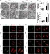
Fatty liver IR aggravates endoplasmic reticulum stress in macrophages and induces mitochondrial calcium overload. Control and HFD-fed mice were treated with a liver IR model or sham procedure. Intrahepatic macrophages were collected at 6 hours after reperfusion. (A) Transmission electron microscopy observe the physical interaction between mitochondria and endoplasmic reticulum in macrophages. (B) The length of the endoplasmic reticulum near mitochondria was measured quantitatively by the normalization of total endoplasmic reticulum length and mitochondrial circumference. (C) Representative fluorescent images of mitochondrial calcium level in macrophages showed green using meg-Fluo4, The mitochondria showed red using mitotracker Red. Control and HFD-fed mice were injected i.p. with TUDCA (400 mg/kg) or PBS (Control), once per day for 3 days before surgery, and then the model of liver IR was established. Intrahepatic macrophages were collected at 6 hours after reperfusion. (D) Representative fluorescent images of mitochondrial calcium level in macrophages showed green using meg-Fluo4. The mitochondria showed red using mitotracker Red. n=4 mice/group. Values were presented as the mean±SD. *p<0.05. Abbreviations: HFD, high-fat diet; IR, ischemia and reperfusion; TUDCA, tauroursodeoxycholic acid.
FIGURE 6
FIGURE 6
Mitochondrial calcium overload aggravates NLRP3 activation by oxidative stress in macrophages. Control and HFD-fed mice were treated with a liver IR model or sham procedure. Intrahepatic macrophages were collected at 6 hours after reperfusion. (A) MDA and GSH/GSSG levels and SOD activity in macrophages. (B) ROS levels were detected by DCFH-DA fluorescence. Control and HFD-fed mice were pretreated with Xestospongin-C (3 μM) or PBS (Control) before ischemia. Intrahepatic macrophages and the liver tissues were collected at 6 hours after reperfusion. (C, D) ROS levels were detected by DCFH-DA fluorescence and 4-HNE. Control and HFD-fed mice were pretreated with mito-TEMPO or PBS (Control) before ischemia. Intrahepatic macrophages were collected at 6 hours after reperfusion. (E) Western blot analysis of NLRP3, C-caspase-1, Pro-caspase-1, IL-1β, IL-18, and GAPDH in isolated macrophages. (F) H&E staining of liver sections. n=4 mice/group. Values were presented as the mean±SD. *p<0.05. Abbreviations: 4-HNE, 4-hydroxynonenal; DCFH-DA, 2,7-dichlorodi-hydrofluorescein diacetate; GAPDH, glyceraldehyde 3-phosphate dehydrogenase; GSH, reduced glutathione; GSSG, oxidized glutathione disulfide; H&E, hematoxylin-eosin; HFD, high-fat diet; IR, ischemia and reperfusion; MDA, malondialdehyde; NLRP3, NOD-like receptor thermal protein domain–associated protein 3; ROS, reactive oxygen species; SOD, superoxide dismutase.
FIGURE 7
FIGURE 7
NLRP3 activation by endoplasmic reticulum stress of macrophages in patients with fatty liver exacerbates IR injury. Liver tissues and peripheral blood were collected respectively before transplantation and before the end of the surgery. (A, B) Serum ALT and AST levels. (C) Gene expression of NLRP3 in liver tissues measured by qRT-PCR. (D) Western blot analysis of P-PERK, IRE1a, ATF6, CHOP, XBP1s and GAPDH in liver tissues. (E) Western blot analysis of NLRP3, C-caspase-1, Pro-caspase-1, IL-1β, IL-18 and GAPDH in liver tissues. n=4/group. Values were presented as the mean±SD. *p<0.05. Abbreviations: ALT, alanine aminotransferase; AST, aspartate aminotransferase; CHOP, C/EBP homologous protein; GAPDH, glyceraldehyde 3-phosphate dehydrogenase; IR, ischemia and reperfusion; NLRP3, NOD-like receptor thermal protein domain–associated protein 3; qRT, quantitative Real-Time.

Similar articles

Cited by

References

    1. Zhai Y, Petrowsky H, Hong JC, Busuttil RW, Kupiec-Weglinski JW. Ischaemia-reperfusion injury in liver transplantation—From bench to bedside. Nat Rev Gastroenterol Hepatol. 2013;10:79–89. - PMC - PubMed
    1. Gao F, Qiu X, Wang K, Shao C, Jin W, Zhang Z, et al. . Targeting the hepatic microenvironment to improve ischemia/reperfusion injury: New insights into the immune and metabolic compartments. Aging Dis. 2022;13:1196–1214. - PMC - PubMed
    1. Cotter TG, Rinella M. Nonalcoholic fatty liver disease 2020: The state of the disease. Gastroenterology. 2020;158:1851–1864. - PubMed
    1. Gedallovich SM, Ladner DP, VanWagner LB. Liver transplantation in the era of non-alcoholic fatty liver disease/metabolic (dysfunction) associated fatty liver disease: The dilemma of the steatotic liver graft on transplantation and recipient survival. Hepatobiliary Surg Nutr. 2022;11:425–429. - PMC - PubMed
    1. Varela AT, Rolo AP, Palmeira CM. Fatty liver and ischemia/reperfusion: Are there drugs able to mitigate injury? Curr Med Chem. 2011;18:4987–5002. - PubMed

Publication types